Southern blotting实验
互联网
PURPOSE:
TolocateaparticularsequenceofDNAwithinacomplexmixture(locateonegenewithinanentiregenome)
SeparatemixtureofDNAsequencesonagel,probewithspecificDNAsequence(gene)
Otherrelatedblottingtechniques:
Westernblotting-identifyasingleproteinwithinacomplexmixture
Separatemixtureofproteinonagel,probewithspecificantibody
Northernblotting-identifyasingleRNAsequencewithinacomplexmixture
SeparatemixtureofRNAsequencesonagel,probewithspecificDNAsequence(gene)
To locate one gene need to cut genome into small, workable pieces using restriction endonuclease digestion
BUT human genome contains 3 billion base pairs of DNA and ~1 million DNA fragments of different lengths can be produced after digestion with 1 restriction enzyme
After restriction enzyme digestion of genomic DNA separate products using gel electrophoresis, get SMEAR of DNA
Southern blot at this point will help identify a specific gene
STEPS
:
1.Cut genomic DNA with restriction enzyme, separate products on agarose gel
DNA must be in denatured state so the probe can bind
the samples you’ll get in lab are single-stranded DNA can use denaturing gel (add SDS to agarose gel)
sometimes scientists soak gel in ~0.5M NaOH, which would separate double-stranded DNA into single-stranded DNA; only ssDNA can transfer onto membrane
A depurination step is optional. Fragments greater than 15 kb are hard to transfer to the blotting membrane. Depurination with HCl (about 0.2M HCl for 15 minutes) takes the purines out, cutting the DNA into smaller fragments. Be aware, however, that the procedure may also be hampered by fragments that are too small.
Neutralize the acid after this step, or the base after the prior step if you don’t depurinate.
2. Transfer DNA fragments from gel to membrane to create a replica (BLOT), membrane usually made of nylon or nitrocellulose
Traditionally, a nitrocellulose membrane is used, although nylon or a positively charged nylon membrane may be used. Nitrocellulose typically has a binding capacity of about 100%26micro;g/cm, while nylon has a binding capacity of about 500 %26micro;g/cm. Many scientists feel nylon is better since it binds more and is less fragile. Transfer is usually done by capillary action, which takes several hours (or minutes in our case!). Capillary action transfer draws the buffer up by capillary action through the gel and into the membrane, which will bind ssDNA.
Sometimes a vacuum blot apparatus is used instead of capillary action. In this procedure, a vacuum sucks buffer (SSC, sodium chloride/citrate) through the membrane. This works similarly to capillary action, except more SSC goes through the gel and membrane, so it is faster (it takes ~1 hour). (SSC provides a high salt level that facilitates transfer of DNA.)
After transferring DNA to the membrane, many scientists treat it with UV light. This cross links (via covalent bonds) the DNA to the membrane.
Nitrocellulose membrane is a high quality membrane ideal for blotting of proteins and nucleic
acids. The nitrocellulose membrane is available in two pore sizes:
0.2 %26micro;m Transfer of low molecular weight proteins (%26lt;20 kDa) and nucleic acids (%26lt;300 bp)
0.45 %26micro;m Transfer of most proteins (%26gt;20 kDa) and nucleic acids (%26gt;300 bp)
Nylon has higher nucleic acid binding capacity and is more durable
3.Incubate membrane with hybridization probe (usually radioactive) that base pairs to specific gene sequence on DNA
Probe the membrane with labeled (radioactive/fluorescent/enzyme) ssDNA. This is also known as hybridization. Process relies on the ssDNA (probe) hybridizing (annealing) to the DNA on the membrane due to the binding of complementary strands.
Probing is often done with 32-P labeled ATP, biotin/streptavidin or a bioluminescent probe.
A prehybridization step is required (usually use gelatin/powdered milk) before hybridization to block non-specific sites, so that the single-stranded probe does not nonspecifically bind anywhere on the membrane.
To hybridize, use the same buffer as for prehybridization, but add the specific probe.
4. Gel position of restriction fragment containing sequences complementary to probe is then detected (usually radioactivity using film)
Visualize radioactively labeled target sequence. If you used a radiolabeled 32-P probe, then you would visualize by autoradiograph. Biotin/streptavidin detection is done by colorimetric methods, and bioluminescent visualization uses luminesence.
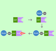
Non-radioactive probes can incorporate biotin (vitamin H)
Detect biotin by its ability to bind to avidin (protein), but avidin NOT colored
Link enzyme that catalyzes a color-producing reaction to avidin
For our experiment - avidin is linked to peroxidase, which is an enzyme that converts chloronapthol to a purple (insoluble) product which allows detection of biotin-labeled DNA (probe)
Our lab:
3 samples to separate on agarose gel
biotin-labeled restriction fragment from bacterial virus bacteriophage lambda calf thymus DNA digested with restriction enzyme BUT NOT biotin-labeled
non-DNA sample
Transfer samples to nylon membrane - not all DNA will transfer, check this by staining gel
DO NOT touch membrane with bare hands - wear gloves
build “sandwich” exactly how it is described in your protocol
smash “sandwich” by placing LARGE book on top
Incubate membrane with TBS-gelatin, gelatin binds all sites on membrane that are not occupied by DNA
Incubate membrane with avidin-peroxidase
Wash membrane
Incubate membrane with color development solution (chloronapthol = substrate)