v468 Chapter 9 自身启动子报告基因验证转录靶点
丁香园
Native Promoter Reporters Validate Transcriptional Targets
Methods in Molecular Biology v468. chapter 9
Otto Schmalhofer, Simone Spaderna ,and Thomas Brabletz
Abstract
The transcriptional activator b-catenin is the key mediator of the canonical Wnt signaling pathway.However, b-catenin does not itself bind DNA, but functions via interaction with T-cell factor (TCF)/lymphoid-enhancing factor (LEF) transcription factors. These proteins contain a high-mobility group(HMG) box that binds DNA in a sequence-specific manner. Thus, in the case of active Wnt signaling,b-catenin activates, in cooperation with proteins of the TCF/LEF family, the expression of a wide variety of genes. To date, the list of established Wnt targets is far from complete. The establishment of plasmidsharbouring reporter genes under control of the native promoter sequences provides a tool to validate novel putative Wnt targets by directly quantifying the b-catenin-dependent activation of each specificgene. In this chapter, we describe how to generate such reporter plasmids using the MMP7 promoteras an example.
Key words: Wnt pathway , Native promoter regions , TCF recognition sites , Reporter plasmids ,Bacmid , Firefly luciferase.
1. Introduction
b-Catenin, the main effector of the canonical Wnt signaling pathway,in cooperation with DNA-binding proteins of the TCF/LEF family, activates the expression of a wide variety of genes (1) .
Numerous direct target genes are already described (a listingis available at: http://www.stanford.edu/~rnusse/pathways/targets.html ). For a more profound understanding of the differenteffects of Wnt signaling, it is necessary to define additional b-catenintargets. A useful strategy to track down putative candidates is thescreening of complementary DNA (cDNA) microarray data, forinstance by comparing data sets of either “normal” cell lines withthe respective cell lines exhibiting an activated Wnt pathway, or tumor cell lines containing stabilized b-catenin with the correspondinglines showing inhibited Wnt signaling (2) . Other possibilitiesare the comparison of tumors with and without CTNNB1mutations (3) or the analysis of tumors with different mutationsand normal tissue (4) (a list of relevant published cDNA arrays isavailable at: http://www.stanford.edu/~rnusse/pathways/array.html ). We use arrays of colon cancer cells transfected with smallinterfering RNA (siRNA) against b-catenin or green fluorescentprotein (GFP) as a control, and arrays of colon carcinomas versusnormal epithelium (unpublished).
First, confirmation of putative targets may be performed viadetection of endogenous protein levels in the respective cell linesor tissue samples, e.g. by Western blot or immunohistochemistry,if appropriate antibodies are available. A method that does notrely on the availability of antibodies is to determine the messengerRNA (mRNA) levels using quantitative reverse transcriptase(RT)-polymerase chain reaction (PCR). The next step after verificationof differential expression levels of the specified mRNA/protein is to validate that the regulation through b-catenin occursat the level of transcription. One way to confirm this is to establishreporter constructs containing the native promoter regionof the putative target gene. Usage of reporter genes, like fireflyluciferase, allows for direct quantification of the promoter activityin response to b-catenin/TCF. This chapter mainly presents thesteps of generating such native reporter constructs , emphasizingthe in silico procedures and theoretical considerations that arenecessary for cloning the reporter plasmids. The exact protocolfor luciferase reporter assays is described in Chapter 8, Volume 1.To demonstrate that activation of reporter genes is directly mediatedvia binding of b-catenin/TCF to its recognition sites withinthe promoter region, additional experiments like electrophoreticmobility shift assays (EMSA) or chromatin immuno precipitations(ChIP) are required.
2. Materials
2.1. Preparation of Bacteria Artificial Chromosome (BAC) DNA
1. LB plates: 10 g Bacto-Tryptone, 5 g Bacto yeast extract, 10g NaCl, 15 g Bacto-agar, and ddH 2 O to 1 L. Autoclave tosterilize, cool to 55°C, add suitable antibiotic, and pour intosterile petri dishes.
2. LB medium: 10 g Bacto-Tryptone, 5 g Bacto-yeast extract,10 g NaCl, and ddH 2 O to 1 L, adjust pH to 7.0 and autoclaveto sterilize, cool to room temperature (RT), and addappropriate antibiotics.
3. Antibiotics for supplementation of LB media (dependent onthe resistance gene encoded by the plasmids/bacmids used):
Ampicillin at 100 μg/mL medium, kanamycin at 25 μg/mLmedium, and chloramphenicol at 12.5 μg/mL medium.
4. Buffer P1, P2, and P3 taken from an arbitrary Qiagen DNAIsolation Kit (Qiagen, Valencia, CA ).
5. Isopropanol.
6. 70% (v/v) ethanol.
2.2. PCR
1. Genomic BAC clone (e.g. from imaGenes, Berlin, Germany).
2. Reporter vector: pGL3basic (Promega, Madison, WI).
3. Specific primers (described in Section 3.2 ).
4. 10 mM dNTP Mix (e.g. from Fermentas, Burlington,Ontario, Canada).
5. 2.5 U/μL Pfu DNA polymerase (e.g. from Fermentas), suppliedwith appropriate 10× reaction buffer.
6. 96% Tetramethylene sulfoxide (TMSO) (e.g. from Sigma,St. Louis, MO).
7. Qiaquick PCR Purification Kit (Qiagen) or similar kit fromother companies.
2.3. Restriction Digest and Ligation
1. Restriction endonucleases (e.g. from Fermentas), theenzymes are supplied with appropriate 10× reactionbuffers.
2. Calf intestine alkaline phosphatase (CIAP; e.g. from Fermentas).
3. 5 U/μL T4 DNA Ligase (e.g. from Fermentas), suppliedwith 10× reaction buffer.
2.4. Transformation
1. Competent bacteria: DH5a subcloning efficiency (e.g. fromInvitrogen, Carlsbad, CA), store at –80°C.
2. SOC Medium: 1 g Bacto-yeast extract, 4 g Bacto-Tryptone,400 μL of 5 M NaCl, 500 μL of 1 M KCl, and ddH 2 O to193 mL, adjust pH to ~7, autoclave to sterilize. All the followingingredients must be sterile filtered and added undera sterile laminar flow hood: 2 mL of 1 M MgCl 2 , 2 mL of1 M MgSO 4 , and 2 mL of 2 M glucose, store these in suitablealiquots of about 5 mL.
2.5. Identification of Clones
1. 10 μM Screening primer forward: 5?-CTAGCAAAATAGGCTGTCCC-3?.
2. 10 μM Screening primer reverse: 5?-CTTTATGTTTTTGGCGTCTTCC-3?.
3. 10 mM dNTP Mix (e.g. from Fermentas).
4. 96% TMSO (e.g. from Sigma).
5. 5 U/μL Platinum Taq Polymerase (e.g. from Invitrogen),the enzyme is supplied with the suitable 10× reaction bufferand MgCl 2 solution (50 mM).
6. Qiaprep Spin Miniprep Kit (Qiagen).
7. Agarose (e.g. from SERVA Electrophoresis, Heidelberg,Germany).
8. 5× TBE-buffer: 54 g Tris base, 27.5 g boric acid, 3.72g EDTA-Na 2 -salt, and ddH 2 O to 1 L (5) , for gel electrophoresis,prepare a 1:10 dilution resulting in 0.5× TBE-buffer.
9. 1-kb Ladder (e.g. from Fermentas).
3. Methods
For validation of putative transcriptional targets of the canonical Wnt pathway via reporter assays, it is necessary to identifythe native promoter region of the respective genes and subsequentlyclone it into a suitable reporter plasmid. Naturally, theexact procedure differs depending on the specific targets to beinvestigated. Additionally, there are of course a variety of differentreporter plasmids available, which may all be more or less useful,depending on the facilities of the specific laboratory. In orderto exemplify the description of the procedure, we have chosenthe published Wnt target MMP7 (6 , 7) for the detailed protocol,including general considerations to be made for each specific targetgene (see Note 1 ).
Concerning the reporter plasmid, we focus on pGL3basic(Promega) harboring the firefly luciferase reporter gene.
3.1. Identification of TCF Sites in Promoter Regions
1. Visit the National Center for Biotechnology Information(NCBI) website at http://www.ncbi.nlm.nih.gov/ .
2. Choose the Gene database from the search box. Enter differentaliases or the gene ID of your gene of interest (GOI), in ourcase MMP7 (gene ID: 4316) in the text box (Fig. 9.1 ).
3. Choose the entry of your species of interest, e.g. homo sapiens(Fig. 9.2 ).
4. On the entrez gene site, click the nucleotide link in theGenomic regions, transcripts, and products text field andchoose the genbank link (Fig. 9.3 ).
5. On the entrez nucleotide site, the sequence of the GOI,MMP7, is displayed. In the header its physical location,nucleotide 101896449 to 101906688 on chromosome 11,is depicted (Fig. 9.4 ).
6. To display the promoter region upstream of the transcriptionstart of the MMP7 gene, add 5,000 bp in the to textbox (see Note 2 ) and click the refresh button (Fig. 9.5 ). Thetranscription start (see Note 3 ) of the GOI now is at position5,001 of the displayed sequence (Fig. 9.6 ).
7. Screen basepairs (bp) 1 to 5,000 of the sequence for theTCF binding sites listed in Table 9.1 .
8. Although there are as many as 14 putative TCF sites in thescreened MMP7 upstream sequence (Fig. 9.7 ), there are onlytwo “classic” sites, as described by Korinek et al. (8) These arelocated in close proximity to the transcription start (indicatedby an arrow in Fig. 9.7 ). Therefore we chose a relatively shortregion of the MMP7 promoter containing these two sites anda third one, as described by Ghiselli et al. (9) , for analysis inreporter assays (see Notes 4 and 5 ).
Fig. 9.3. MMP7 gene entry at the Entrez Gene site.
Fig. 9.6. Position of mRNA in the displayed sequence.
Fig. 9.7. Putative TCF binding sites in the MMP7 promoter region.
Table 9.1
Pipetting instructions for PCR amplification of promoter sequences
3.2. Selection of Primer
1. For cloning this fragment by means of a traditional strategy intothe reporter plasmid pGL3-Basic from Promega (Fig. 9.8 ),choose restriction enzymes from the vector’s multiple cloningsite (MCS) that do not cut within the region of interest(see Note 6 ). Use one of the numerous online tools, e.g. theNew England Biolabs (NEB) cutter at the NEB homepage athttp://tools.neb.com/NEBcutter2/index.php .
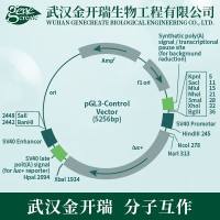
Fig. 9.8. pGL3 Basic vector from Promega.
2. Paste the sequence from bp –1,000 to +48 relative to thetranscriptional start site of the MMP7 gene in the sequencewindow and submit (Fig. 9.9 ).
Fig. 9.9. Homepage of NEBcutter 2.0.
3. Xho I and Hin dIII from the the MCS of pGL3-Basic do notcut within this region, as you can learn from the 0-cutter list(Fig. 9.10 ).
Fig. 9.10. Display of restriction analysis at the NEBcutter 2.0 site.
4. For selection of primers, use one of the various tools availableonline, e.g. OligoPerfect TM Designer from Invitrogenat http://www.invitrogen.com/content.cfm?pageid=9716 .Paste the sequence of interest into the Target Sequence textbox, select PCR:Cloning from the application box, and submit(Fig. 9.11 ).
Fig. 9.11. OligoPerfect TM Designer at the Invitrogen homepage.
5. A suitable region of the MMP7 promoter to amplify is bp–844 to +35 (relative to the transcription start site), containingthe three identified TCF sites. Add the recognitionsequences of Xho I and Hin dIII and choose TraditionalCloning (Fig. 9.12 ).
6. The Oligo Perfect TM Designer delivers different primer pairs(Fig. 9.13 ). For example, as forward primer 5¢-acca ctcgag tggatctccaagttgaaggtc-3¢ and as reverse primer 5¢-atgc aagctt -gagacaattgttcttggacctatg-3¢. Underlined are the recognitionsequences for Xho I or Hin dIII, respectively. Highlighted areadditional oligonucleotides, added 5¢ for improving restrictionefficiency.
Fig. 9.13. Cloning primer search results for the MMP7 promoter by Oligo Perfect TM Designer.
3.3. Selection and Preparation of Bacmid
1. For identification of a BAC containing the genomic locus ofhuman MMP7 for use as template in PCR (see Note 7 ), visit thewebsite of imaGenes, formerly the German Resource Centerfor Genome Research, at http://www.imagenes-bio.de/and choose Product Search .
2. Choose the species and the type of DNA clone of interest,which is homo sapiens and genomic clone, and search forMMP7 (Fig. 9.14 ).
Fig. 9.14. Search options of the GenomeCube.
3. Order the listed genomic DNA clone RZPDB737H032090D(Fig. 9.15 and Note 8 ).
Fig. 9.15. MMP7 search results with the GenomeCube.
4. Preparation of Bacmid DNA is modified from Sambrooket al. (5) and closely follows instructions of the Qiagen miniprepmanuals. Buffers from Qiagen are used for isolation.The yielded DNA is of poor purity; however, the quality issufficient for use in subsequent PCR.
5. Pick a clone from a freshly streaked selective plate and incubateovernight at 37°C in 3 mL LB with the appropriateantibiotic on a shaking platform.
6. Harvest bacteria by centrifugation in a tabletop centrifugefor 5 min at 6,000 × g .
7. Discard supernatant and resuspend pellet with 100 μL buffer P1.
8. Add 100 μL buffer P2, mix by gently inverting, and incubatefor 5 min at RT.
9. Add 100 μL buffer P3, mix by gently inverting, and incubatefor 10 min on ice.
10. Centrifuge with full speed in a microfuge (~17,900× g ) for10 min at RT.
11. Transfer the supernatant to a new microtube and add 200μL isopropanol.
12. Centrifuge with full speed in a microfuge (~17,900× g ) for15 min at RT.
13. Discard the supernatant; add 200 μL of 70% ethanol andcentrifuge for another 5 min.
14. Discard the supernatant and air-dry the pellet for 5–10 min(see Note 9 ).
15. Resuspend the DNA pellet by adding 50 μL H 2 O. Determineand adjust the concentration to 100 ng/μL.
3.4. PCR
1. For amplification of the selected promoter fragment, preparethe PCR reaction mix according to Table 9.2 (see Note 10 ).
2. Use the thermocycling program according to Table 9.3 .
3. Apply 5 μL of the reaction mix to electrophoresis on a 1%(w/v) agarose gel to check for correct size of the amplicon.
4. For subsequent purification of the amplicon from the reactionmix, use the Qiagen PCR Purification Kit according tothe user manual, and elute in 30 μL of H 2 O.
3.5. Digest and Ligation
1. For digestion of the pGL3-Basic vector and the PCR amplicon,prepare the following reaction mixtures according toTable 9.4 and incubate overnight at 37°C.
2. Incubate for 20 min at 80°C to heat-inactivate restrictionenzymes.
3. For dephosphorylation of the linearized pGL3-Basic, add1 μL of CIAP and incubate at 37°C for 30 min. InactivateCIAP by incubating at 85°C for another 15 min.
4. For subsequent purification of digested vector and ampliconfrom the reaction mix, use the Qiagen PCR Purification Kitaccording to the user manual.
5. For ligation, prepare the following reaction mix accordingto Table 9.5 , including a re-ligation reaction of the vectoras a control. Incubate for 2 h at RT or overnight at 16°C (seeNote 11 ).
Table 9.2
Cycling conditions for PCR amplification of promoter sequences
Table 9.3
Pipetting instructions for endonuclease digestion
Table 9.4
Pipetting instructions for endonuclease digestion
Table 9.5
Pipetting instructions for DNA ligation Ligation volume Religation
3.6. Transformation
1. Thaw bacteria (DH5α subcloning efficiency) on ice.
2. Transfer 50 μL of bacteria in a pre-chilled reaction tube.Then add 5 μL of the ligation mix and resuspend gently.
3. Incubate on ice for 30 min.
4. Heat shock for 20 sec at 37°C.
5. Incubate on ice for 2 min.
6. Add 500 μL SOC medium and incubate on a shaker at 300rpm for 1 h at 37°C.
7. Centrifuge 30 sec with full speed in a tabletop microfuge.Discard 400 μL of the supernatant.
8. Resuspend the pellet in the remaining supernatant andstreak on a selective plate containing ampicillin.
3.7. Identification of Clones
1. For identification of bacterial clones that carry the pGL3-Basicvector with the correctly inserted amplicon, prepare a PCRmastermix for as many bacterial clones as you intend to screenaccording to Table 9.6 . Prepare two additional reactions fornegative and positive controls. Transfer 25 μL of the mix to aPCR tube, pick a bacterial clone from the selective plate, streakthe clone on a masterplate, and resuspend it in the PCR tube. Aspositive control, pick a bacterial clone from the re-ligation plate.
2. Use the thermocycling program according to Table 9.7 .
3. Subject 5 μL of each PCR to agarose gel electrophoresis.
Clones that did not incorporate the insert will produce anamplicon of 170 bp, e.g. the positive control clone, whereasclones carrying the correct insert from the MMP7 promoterwill produce an amplicon of 1,028 bp (see Note 12 ).
4. Prepare plasmid DNA from a positive clone from the masterplateby using the miniprep kit from Qiagen and subject it tosequencing reaction. When the region of interest is insertedcorrectly, perform luciferase assay analysis (see Chapter 8)(Volume 1, Biechele and Moon).
Table 9.6
Pipetting instructions for screening PCR
Table 9.7
Cycling conditions for screening PCR
4. Notes
1. Mine the PubMed database for the gene of interest and if thereare published results on the promoter region and a reporterplasmid is already described, request it from the author.
2. Depending on the localization of the coding sequence of theGOI on the plus or minus strand of DNA, withdraw 5,000bp in the from text box or add 5,000 bp in the to text box,respectively, for investigation of the 5¢ region.
3. Be mindful to check your GOI for splice variants displayingdifferent transcription start sites and thus different upstreamregulatory sequences.
4. Be mindful that in most publications on b–catenin/TCF targetgenes, the prevalent TCF site identified is the WWCAAAGsequence; there are many fewer publications on the otherTCF sites listed in PubMed. Be aware that additional TCFsites might be identified.
5. Depending on the localization and frequency of TCF sitesin your specific 5¢ region, you may decide to clone a shorterand a longer region of the promoter.
6. For simplicity of the subsequent cloning procedure, alwaystry to choose restriction enzymes that work with high efficiencyin a double digest.
7. Genomic DNA may also be used as a template; however,efficiency of the PCR strongly improves when a BAC is usedas template.
8. When clicking on the clone designation button, you getadditional data, including the international clonename (e.g.RP11-315O6). Using this name, you should be able to orderthe clone from different distributors (e.g. BACPAC Resourcesat CHORI Oakland, CA; http://bacpac.chori.org ).
9. If the pellet is dried for too long, it becomes insoluble.
10. When cloning promoter regions, one often has to deal withGC-rich sequences, so addition of TMSO (10) or other reagentssuch as dimethylsulfoxide (DMSO) or Betaine usuallystrongly improves PCR efficiency. Use a proofreadingpolymerase, e.g. Pfu polymerase from Fermentas, to ensurecorrect amplification of the target sequence .
11. One may also try different ratios of Insert/Vector in the ligationreaction mix. In the case of digestion with blunt-end-generatingrestriction enzymes, e.g. Sma I, use polyethylene glycol (PEG),which is supplied with T4 ligase from Fermentas, accordingto the user manual, to increase ligation efficiency.
12. If it was difficult to amplify the region of interest in the firstplace, the screening PCRs may not produce an amplicon,except for the positive control. Nevertheless, the screenedclones may carry the insert. It is reasonable to characterizethem further by performing minipreps and subsequentrestriction controls.
Acknowledgments
This work was supported by grants to TB from the GermanResearch Ministry BMBF (NGFN2 project no. 01GS0436). SSwas funded by the German Research Council DFG (grant no.BR1399/4-3) and OS was funded by the Deutsche Krebshilfe(grant no. 106958).
References
1. Giles, R.H., van Es, J.H., Clevers, H. (2003)Caught up in a Wnt storm: Wnt signaling in cancer. Biochim Biophys Acta 1653 , 1–24.
2. van de Wetering, M., Sancho, E., Verweij, C., de Lau, W., Oving, I., Hurlstone, A., van der Horn, K., Batlle, E., Coudreuse, D., Haramis, A.P., Tjon-Pon-Fong, M., Moerer, P., van den Born, M., Soete, G., Pals, S., Eilers, M., Medema, R., Clevers, H. (2002) The beta-catenin/TCF-4 complex imposes a crypt progenitor phenotype on colorectal cancer cells. Cell 111 , 241–250.
3. Zirn, B., Samans, B., Wittmann, S., Pietsch,T., Leuschner, I., Graf, N., Gessler, M. (2006) Target genes of the WNT/betacateninpathway in Wilms tumors. Genes Chromosomes Cancer 45 , 565–574.
4. Huang, S., Li, Y., Chen, Y., Podsypanina, K., Chamorro, M., Olshen, A.B., Desai, K.V., Tann, A., Petersen, D., Green, J.E., Varmus, H.E. (2005) Changes in gene expression during the development of mammary tumors in MMTV-Wnt-1 transgenic mice. Genome Biol 6 , R84.
5. Sambrook, J., Fritsch, E.F., Maniatis, T. (1989) Molecular cloning: a laboratory manual. 2nd edn. Cold Spring Harbor LaboratoryPress.
6. Brabletz, T., Jung, A., Dag, S., Hlubek, F.,Kirchner, T. (1999) beta-catenin regulates the expression of the matrix metalloproteinase-7 in human colorectal cancer. Amer J Pathol 155 , 1033–1038.
7. Crawford, H.C., Fingleton, B.M., Rudolph-Owen, L.A., Goss, K.J., Rubinfeld, B., Polakis, P., Matrisian, L.M. (1999) The metalloproteinase matrilysin is a target of beta-catenin transactivation in intestinal tumors. Oncogene 18 , 2883–2891.
8. Korinek, V., Barker, N., Morin, P.J., van Wichen, D., de Weger, R., Kinzler, K.W., Vogelstein, B., Clevers, H. (1997) Constitutivetranscriptional activation by a beta-catenin-Tcf complex in APC-/- colon carcinoma. Science 275 , 1784–1787.
9. Ghiselli, G., Coffee, N., Munnery, C.E., Koratkar, R., Siracusa, L.D. (2003) The cohesin SMC3 is a target for beta-catenin/ TCF4 transactivation pathway. J Biol Chem 278 , 20259–20267.
10. Chakrabarti, R., Schutt, C.E. (2002) Novelsulfoxides facilitate GC-rich template amplification. Biotechniques 32 , 866, 868, 870– 862, 874.
11. Rockman, S.P., Currie, S.A., Ciavarella, M.,Vincan, E., Dow, C., Thomas, R.J., Phillips, W.A. (2001) Id2 is a target of the beta-catenin/T cell factor pathway in colon carcinoma. J Biol Chem 276 , 45113–45119.
12. Gradl, D., Kuhl, M., Wedlich, D. (1999) The Wnt/Wg signal transducer beta-catenin controls fibronectin expression. Mol Cell Biol 19 , 5576–5587.