(共享)ELISA原理与实验方法
丁香园论坛
ELISA原理与实验方法
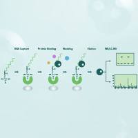
The advantages of the ELISA are similar to other antibody-labeled reactions which include specificity, sensitivity, inexpensiveness, and safety. Since the enzyme label is the critical portion of ELISA, its selection is very important. The enzyme selected should be stable under the conditions used for storage, cross-linking, and the assay. The most effective enzymes will have a high specific activity and will be inexpensive. Equally important is that the enzyme must be absent from the antigen or the antiserum preparations used in the serological tests, otherwise false positive tests would result. False negative results could stem from the presence of enzyme inhibitors or inactivators in the serological reagents. Appropriate controls must be incorporates into the test to identify these potential problems in ELISA. The enzyme catalyzes a change in a substrate molecule, but the enzyme itself is not consumed in the process. The enzyme molecule continues to act on more substrate molecules and produce thousands of colored products. Thus, in ELISA, antigens are detectable when present in only nanogram and picogram quantities. One form of the ELISA is carried out in a microtiter plate which is used as the solid phase for adsorption of antigen (Figure 1). Albumin or powdered milk is used to block the remaining sites which could result in false positives. Monoclonal antibodies will react with the antigen attached to the microtiter plate. The complex antigen-antibody is then reacted with enzyme conjugates to a goat antimouse IgG, which catalyzes the substrate p-nitrophenyl phosphate, to form a colored product. Excess reagents are removed by washing, and enzyme activity can be measured with an ELISA plate reader at 415nm. The amount of colored product is an indirect expression of the monoclonal antibody amount in the sample.
Methods
1 Obtain a 96-well plate and rinse 3x with PBS.
2 Add 50 µl of a 1 mg/ml of antigen (venom, serum, or fraction), incubate for 30 min at 37°C.
3 Rinse with PBS 3x.
4 Add 5% powdered milk, incubate for 30 min at 37°C.
( 5g powdered milk in 95 ml PBS)
5 Rinse with PBS 3x.
6 Add 50 µl of antibody, incubate 30 min at 37°C.
7 Rinse with PBS 3x.
8 Add 50 µl of goat anti-mouse IgG conjugate with alkaline phosphatase.
(1 µl of conjugate in 4 ml of PBS)
9 Incubate 30 min at 37°C.
10 Rinse 3x with 0.05% Tween 80 in 0.01 M PBS, pH 7.4.
11 Rinse 3x with HPLC water.
12 Add 50 µl of color development reagent
(1 tablet of p-nitrophenyl reagent in 5 ml of 10% diethanolamine, pH 9.8) (mix well)
13 Incubate 30 min at 37°C.
14 Just to check enzyme and color product put 100 µl of each in a test tube and watch for a yellow color change.
Figure 1. Schematic of an ELISA
MATERIALS
Phosphate-buffered saline (PBS) ,0.01 M Na2HPO4,0.15 M MaCl, PH 7.4
Sodium phosphate dibasic (Na2HPO4) 0.87 g
Sodium phosphate monobasic (Na2HPOundefined H2O) 0.54 g
Sodium chloride 8.5 g
HPLC water 1.00 L
Adgust PH to 7.4 with 1N NaOH 1M HCl
SLGMA Immunp Chemical SIGMA Diagnostics
Anti-mouse IgG (Fab specific) Sigma 104 Phosphatase
Alaline Phosphatase conjugate Substrate Tablete
(antibody developed in goat) St. Louis,Mo 63178, USA
Affinity isolated antibody adsorbed Lot # 043H6178
With bovine horse and human serum protein Cat # 0h176
Lot # 055H4835
Lot # A-2179
Tween 80
Laboratories ,Detroit,Michigan ,USA
1 drop of Tween 80
150ml of PBS , 0.01 M, PH 7.4
Reference/More information
Voller A , Bartlett A, Bidwell DE, Clark MF, Adams An, 1976. The detection of viruses by enzyme-linked immunosorbent assay (ELISA). J Gen Virol 33, 165-167.
Figure 1