vol 687 Chapter 1 PCR实验室建设
丁香园
5849
好多同学在做PCR时,都会面临一个问题——污染,本期师兄通过引用著名的Metods系列,告诉怎么有效减少PCR的污染问题。
想跟师兄探讨的,扫描下方的二维码向我申请(微信号:shixiongcoming),申请理由注明:PCR +你的专业+学校或单位

开启PCR英语时间
Setup of a PCR Laboratory
Methods in Molecular Biology vol.687 Park D.J. (ed.) PCR Protocols Chapter 1
Abstract
PCR represents an extremely powerful and central molecular biology method. At the heart of its power is the exquisite sensitivity offered: single molecule detection in certain contexts. However, with greatpower comes great responsibility. Contamination of reagents or test samples with amplifiable material,such as previous reaction products, can be crippling to scientists applying PCR protocols. Prevention of PCR contamination is far and away preferred over eradication. This chapter sets out to offer guidance as to how to use PCR while minimising contamination problems.
Key words: PCR laboratory setup, PCR contamination, PCR reagents, PCR equipment
1. Introduction
From its beginnings in 1985, PCR technology has become acornerstone of molecular biology. As PCR became more widelyused, scientists discovered its strengths and weaknesses. Amplification to detectable levels can be achieved from even singlecopy of template nucleic acids. PCR can be used to amplifyfrom not only DNA but also RNA and the products can now bedetected in real time. However, the attribute that makes it mostappealing is the same attribute that is the source of much frustration. PCR contamination, once established, can be very difficult to rid oneself of. The best approach is to prevent PCR contamination from happening in the first instance. With a good laboratory setup and good technique, many ofthe problems associated with PCR can be avoided. However, onemust always remember that all we can do is to reduce the risk toacceptable levels. The more procedures we implement, the furtherreduced are the risks of PCR contamination. This must be balanced with creating a work environment that is comfortable towork in. Implementing too many additional procedures mayresult in a tendency to not follow what are perceived as unnecessaryprocedures.
The appropriate balance between ease of work and preventingPCR contamination will be different for different organisations. A laboratory manufacturing PCR diagnostics kits toregulatory standards would need to take the most stringent measuresto prevent PCR contamination, whereas a laboratory whichperforms PCR infrequently for research purposes may not needsuch stringent controls, and the balance would be tipped moretoward ease of work. These are decisions which, ultimately, eachlaboratory must make based on their requirements.
This chapter outlines some general considerations for setting up a PCR laboratory with a view to preventing contamination.
2. Materials
Materials should be stored as specified by the manufacturer.
Where a material is made within the laboratory, all bottles shouldbe clearly and uniquely labelled with at least the manufacturer’sinitials or name, name of the solution or the material, and theexpiry date. Materials should be placed in the rooms they are tobe used in, and where one material is required in multiple rooms,each room should have a dedicated stock. Materials should not bemoved from higher copy to lower copy rooms as this can increasethe risk of transferring contamination between rooms. If necessary,but as a last resort, procedures should be implemented todecontaminate materials (see Note 1). In case of laboratory contamination,some guidelines are provided in Subheading 3.
There are many different pieces of equipment which areimportant for a PCR laboratory. Later, the placement of criticalequipment is specified, but here some of the more importantpieces of equipment are introduced.
1. Fridges and freezers are used for storing reagents andsamples. Generally, a fridge or freezer should display thetemperature or contain probes for monitoring temperature. Temperature should be monitored regularly such that anyvariations of temperature outside specification can berecorded. Preferably an alarm should be connected. It is alsoimportant to know for how long a fridge or freezer hasbeen out of specification. Freezers are typically available aseither -20 or -80°C models. A -80°C freezer is recommendedfor long-term storage of nucleic acids and a -20°Cfreezer is adequate for shorter periods of storage and isgenerally used to store PCR reagents in common use (1). Ideally, calibration should be performed annually. Frost- free freezers should be avoided for the storage of molecularbiology reagents (see Note 2).
2. Pipettes are fundamental tools within the laboratory. Theycome as single channel, multichannel, and/or stepper/repeater pipettes. They are available as positive-displacementor air-displacement models. Positive-displacement pipettesare more accurate for pipetting viscous liquids (2) and whenused appropriately can help to prevent contamination as theentire tip and mechanism is ejected after use. Use of barriertips with air-displacement pipettes is strongly recommendedto reduce contamination derived from PCR products or othersources via aerosols (3). Calibration of pipettes is critical toensure accurate and reproducible results. Currently, there areno definitive regulations on how often calibration shouldoccur. However, the Center for Drug Evaluation and Research,FDA, Guidance for Industry, Subpart D, Equipment states“the key point is that the calibration schedule should be frequentenough to assure data validity...”. Factors which affect howoften calibration should occur include:
●● The liquid that is dispensed. Corrosive liquids can formvapours which can damage the internal mechanisms of thepipette.
●● Handling of pipettes. Pipettes that are often dropped aremore subject to wear and tear, and will require more frequentcalibrations.
●● The accuracy required. Facilities that require high accuracyin their procedures need to regularly monitor the adequacyof their pipettes.
In general terms, the author recommends that most researchlaboratories should calibrate pipettes every 6 months. Facilitieswhich operate under GMP regulations should consider calibrationevery 3 months.
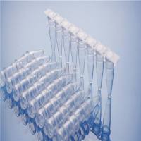
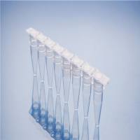
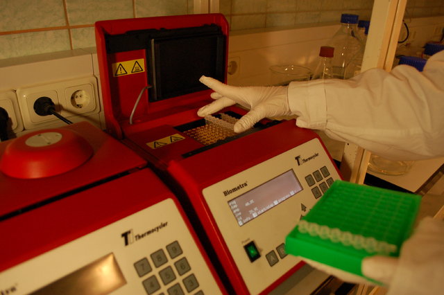
3. Thermocyclers are required to perform PCR. There are manymanufacturers making a variety of models. Thermocyclersinclude machines with 96- or 384-well format blocks. Somehave interchangeable blocks. Reactions can occur in singletube, strips, 96- or 384-well plates, or capillaries. Reactionvessel volumes for use in conventional PCR are typically 0.2 or0.5 mL. Ramp rates and uniform heat distribution are importantfactors influencing the consistency of operation. It is recommendedthat these be calibrated on a regular basis. Whenutilising several different brands of PCR machines, one shouldnot assume that they will perform identically. Particular attentionshould be paid to the ramp rates, tube size, and block architecture. Real-time PCR technology allows amplificationand analysis to take place simultaneously without the need forfurther handling. This reduces PCR contamination risk as thecompleted reaction can be discarded immediately after processingwithout exposure to the laboratory environment.
4. Cleaning with 2–6% hypochlorite (w/v) has been shown todegrade DNA (4) (see Note 3). At this concentration, thelevels of hypochlorite are similar to household bleach; alwaysrefer to the manufacturer’s MSDS for safety and disposalinstructions. Regular cleaning of surface area before and afterwork can reduce the spread of DNA and prevent contamination.
A value of 70% (v/v) ethanol is often used to wipebenches after hypochlorite treatment to prevent it from interferingwith PCRs. Regularly changed bench liners and a regularlaundry schedule are important elements.
5. Planning a PCR laboratory: the most common setup for a PCRlaboratory involves three separate areas. It is highly recommendedthat at least three separate rooms should be used forthe three major steps of PCR: the Master Mix preparation andpre-PCR reagent handling and addition, the sample addition,and the PCR and analysis. Implementing such physical separationbetween the three steps reduces the risk of products contaminatingreagents or samples to be included in future PCRs.
These areas are generally termed as a Pre-PCR Room, a LowCopy Room, and a High Copy Room. Work flow should occur ina unidirectional manner (Fig. 1), and at no point should samples or equipment from downstream, including laboratory coats andgloves, be transferred to upstream areas. Each room should havedesignated equipment and instruments which should not bemoved between rooms. Ideally, the Pre-PCR Room and LowCopy Room should be positively pressurised relative to thecorridor.
The High Copy Room should be negatively pressurisedrelative to the corridor. In this manner, PCR aerosols or contaminateddust are discouraged from entering the Low Copy andPre- PCR areas (see Note 4). Where such physical separation isnot possible, three separate areas should be established. If onlytwo rooms are available, the low copy area and the Pre-PCR areacan be shared. Separate equipment and instruments should bearranged for each area and transfer of equipment and instrumentsbetween areas should be avoided. For the pre-PCR setup, a laminarflow hood should be used.

Fig. 1. Recommended workflow through a PCR laboratory. Workflow should always beinitiated in the pre-PCR area and flow progressively through the other work areas. Theflow is unidirectional and materials should never flow backwards.
3. Methods
1. Routinely one must gown up and glove up upon enteringthe room and degown and remove gloves just before leavingthe room (see Note 5). This is good practise for all laboratoryareas and ensures that whatever work has been done inthat area stays in that area and does not travel to otherareas.
2. Use of bench liners will contain any spills that may interferewith the PCR; these should be regularly changed and monitored. Any spills should generally be wiped using absorbentdisposable wipes. When wiping spills, do not smear the surface,but absorb the spill and dispose of the wipe.
3. Regular cleaning of surfaces with 2–6% hypochlorite (w/v),followed by 70% (v/v) ethanol ensures a good clean environmentto work in (5). Along with maintaining a clean environment,one must always be conscious of spreading
contamination. Contaminated bench tops and door handlesare major risks (see Note 6). Regular changing of gloves canprevent such spread. It is further recommended that glovesbe removed immediately prior to exiting a room (seeSubheading 3, step 1); gloved hands should not come intocontact with door handles as this can easily spread contaminationbetween laboratory areas.
4. Always centrifuge samples before opening as these can be asource of aerosols which can contaminate laboratory areas.
5. Gel areas are a potent source of contamination. Gel areasmust always be located in the High Copy area and where possibleshould be segregated from the rest of the laboratory Particular care must be taken with buffers such as those usedduring electrophoresis, as PCR products have been in directcontact with the solutions. Always change gloves before leavingthe gel area.
6. All equipment should be dedicated for each area and shouldnot be moved between areas. Equipment should be regularlymonitored for performance and calibrated as required.
7. The Pre-PCR Room is routinely used for preparation ofMaster Mixes and storage of pre-PCR reagents. It is criticalthat this area be kept free of any DNA or RNA, especiallythose related to work routinely performed in the laboratory. Any contamination here can be hard to identify, time-consumingto investigate, and expensive to remedy. General guidelineson how to deal with established PCR contamination areincluded later in this chapter. One must always be very cautiouswhen bringing material into the room. The Pre-PCRRoom should contain everything that is required within theroom such that there is no need for laboratory personnelto exit and re-enter the room during pre-PCR processing. The Pre-PCR Room should contain a ?20 or ?80°C freezerfor storage of PCR reagents and a vortex for mixing reagents;however, enzymes or enzyme mixes should not be vortexed. The Pre-PCR Room should also contain a microfuge to“pulse down” reagents following mixing (see Note 7). Alwayscheck to ensure no liquid is present in the lid as this can dropout and contaminate bench surfaces and gloves. Bench surfacesshould be regularly cleaned before and after setup using2–6% hypochlorite (w/v), followed by 70% (v/v) ethanol. Dedicated pipettes and tips for liquid handling should be used.A laminar flow cabinet is highly recommended for preparingMaster Mixes. If laminar flow cabinets cannot be utilised,special PCR cabinets can be used. Many cabinets comeequipped with UV lights (see Note 8). These should beturned on before and after PCR setup for at least 30 min. UVradiation can also be used to remove contaminations fromPCR Master Mixes for use in the PCR. Studies have shownthat exposure to UV light for a period of 30 min can inactivatemany DNA species in solution by approximately fiveorders of magnitude (6) (see Note 9). Other equipment usedin preparation can also be routinely decontaminated using UVlight. It is suggested that this be done in conjunction withbleach and ethanol cleaning on a regular basis as part of thelaboratory cleaning schedule.
8. The Low Copy Room is often used for the preparation, storage,and addition to reaction mixtures of DNA or RNA samplesused in PCR. No PCR product or DNA clones containingtarget sequences should be handled in this area. This area should contain a ?20 or ?80°C freezer for storage of DNA,vortex mixers, and centrifuges. Consideration should be givento how much freezer space is required; in some cases, thelaboratory may desire to store samples for a number of years,in which case provisions should be made to ensure there isadequate space for storage (see Note 10). Samples such asblood, serum, or cultured cells can be brought into this roomfor the purpose of extracting DNA or RNA, assuming appropriatesafety precautions are in place. If working with infectiousmaterials, ensure that appropriate systems and equipmentare in place for the protection of both laboratory staff andsamples (see Note 11). It is ultimately the responsibility ofthe laboratory head and senior staff to comply with any biosafetyregulations applicable to their laboratory. Dedicatedtools and equipment should be utilised in this room. Use ofbarrier tips or positive-displacement pipettes discourages thespread of aerosols and should be used throughout the PCRprocedure. To further reduce the spread of aerosols and liquidcollected around vessel lids, one should always centrifugesamples briefly before opening. Use of screw cap tubes ispreferred over “popping” lids as the latter can increase theprobability of aerosol formation. When extracting DNA orRNA, a method that requires the least amount of handling isrecommended. This not only decreases the risk of spreadinggenetic material, but also reduces the risk of contaminationbetween samples. For large numbers of extractions, automatedsystems provide a secure, contained environmentwhere DNA or RNA extractions can occur with a reducedrisk of spreading material throughout the laboratory. Theclosed space allows for quick and easy decontaminationbetween runs. Automated DNA extraction systems providereproducible and reliable quantities and qualities of DNAwhich are not subject to the variation which can occur whenhuman operators are involved. They reduce the possibility ofsample mix-up which is a very important consideration fordiagnostic laboratories. A disadvantage of these systems canbe their reduced sensitivity with some samples (7).
9. The High Copy Room is so termed because of the presenceof high copies of DNA and PCR products. This area is usedfor the amplification of low copies of DNA to yield high copiesof DNA. A PCR mix (post-template addition) is broughtinto this area and amplified using a thermocycler. The productsgenerated by PCR are a major source of contaminationwithin the laboratory. This contamination principally occursfrom aerosols which arise from the process of pipetting andmanipulating the DNA, contaminated liquids being splashed,and surface-to-surface contact. Several strategies have been introduced to minimise this contamination; the same methodscan be utilised in this area. These contaminants cause fewproblems if confined to the High Copy Room but can causehavoc if allowed to spread to the other areas. It is thereforecritical that no mixing of reagents and equipment be allowedbetween this area and the other areas of the laboratory. Thisarea contains much of the equipment, such as thermocyclers,centrifuges, qPCR facilities, sequencing facilities, electrophoresisequipment, and imaging equipment including transilluminatorsand cameras for photographing gels. It also oftenserves as the storage site for PCR products. Although PCRproducts can be stored in this area they must not be taken outof this area unless being disposed of in a suitably containedmanner.
10. Discard: all items for disposal in the High Copy Room aretransferred only when appropriately contained to preventexposure of other areas of the laboratory to high copy material. Waste contract services are recommended as they willsupply and control the management of waste generated bythe laboratory. There are three major classifications of hazardouswaste (8) routinely generated during normal PCRlaboratory functioning. The first is infectious waste/highlyinfectious waste which is routinely used for the disposal ofPCR products, contaminated agar plates, live cultures, humancells and blood, and disposables that have been in contactwith the above. The second is sharps which includes objectsor devices that have acute, rigid corners, edges, points or protuberancescapable of cutting or penetrating the skin, e.g. hypodermic needles, glass, scalpel blades, and lancets. Allsharps are hazardous because of the potential to cause cutsand punctures. Sharps may also be contaminated with toxic,infectious, or radioactive materials, which would increasesubstantially their potential to cause harm. Thirdly, there ishazardous chemical waste. Chemical waste consists of discardedchemicals (solid, liquid, or gaseous) that are generatedduring disinfecting procedures or cleaning processes, forexample. They may be hazardous (toxic, corrosive, and flammable)and must be used and disposed of according to thespecification formulated on each container. Nevertheless nonexplosiveresidues or small quantities of outdated productsmay be treated together with infectious waste.
11. In cases where the above measures relating to physical separationare impossible, or as an additional measure, “temporal” separation of areas can be employed. This separates work tasksin time such that low copy and high copy materials are neverhandled at the same time. Combined with appropriate cleaningschedules and the use of separate equipment, consumables, and areas, this approach can help to reduce the risk ofintroducingPCR contamination into reagent stocks or templatematerial (see Note 12).
12. Enzymatic treatment of PCR Master Mix is an elegant,optional measure to help to limit “PCR carryover” (9).However, this should not be viewed as a replacement for anyof the other measures discussed in this chapter (see Note 13).
The basic principle involves the substitution of dTTP withdUTP, where dUTP is used as substrate during polymerisationcatalysed by compatible polymerases. The PCR productformed after this substitution is susceptible to enzymaticcleavage. Incubation of PCR Mix with a quantity of uracil-DNA N-glycosylase before addition of any template removesPCR product “carry over” that may have found its way intothe Master Mix. Subsequent heating at 90°C destroys theenzyme. Many kits are available to streamline this process andin order for this method to be effective it must be incorporatedas early as possible. UV irradiation of the PCR MasterMix is another option which can be used to limit PCR carryover(10) (see Note 9).
13. Negative controls must always be employed so that anycontamination can be detected as early as possible. When contaminationis found, a pre-planned strategy must be put intoaction as quickly as possible.
14. If PCR contamination is detected in the laboratory, one oftwo approaches can be used. The first approach centres onthe quick eradication of PCR contamination. This approachwill quickly remove the source of the contamination but canbe very expensive. It will not identify the source of the contamination. In this approach, the laboratory must discard allreagents used in PCR preparation, including primers, buffers,dNTPs, water, and enzymes. Also included can be the discardingof equipment and consumables such as pipettes andPCR plates or tubes. Thorough decontamination of workspacesand any equipment that cannot be discarded should beperformed. The second approach involves the testing of allreagents and equipment. This can be a very time-consumingexercise, but the source of the contamination can be isolated. It can be less expensive (depending on the resources consumedin terms of personnel-time, reagents consumed, andultimate success of the process) as only the source of the contaminationneed be discarded and measures can be put in place to prevent future contaminations of the identified component. In this approach, fresh reagents are prepared fromknown PCR-clean sources. Each suspected reagent is testedin isolation to identify which component is the source of thecontamination.
15. Although the utilisation of the aforementioned techniqueswill never ensure the prevention of PCR contamination, theywill reduce the risk of PCR contamination. By thinking of allpossibilities and planning the work environment, we can substantiallyreduce the risk of contamination occurring.
16. Good training of laboratory staff is essential regardless of howwell planned a laboratory is. Staff should understand the reasonsfor the systems that are in place and the cost of havingPCR contamination within the laboratory (see Note 14). Formal laboratory training of all staffs in laboratory protocolsand procedures should be accompanied by signed trainingforms and regular reviews to ensure compliance. The laboratorystaff will ultimately determine how the laboratory functions,and as such, the resources required for them to be ableto perform their duties efficiently and effectively should bemade available to them.
4. Notes
1. Decontamination should be viewed as a last resort. Preventionof PCR contamination is by far the preferred alternative.
2. Frost-free freezers cycle through rounds of relatively warmerand cooler temperature cycles. This results in multiple freeze–thaw events which can be damaging to the stored PCR reagentor template nucleic acid.
3. Hypochlorite bleach must be prepared relatively fresh and bewithin specification. Active species degrade over time to resultin reduced bleaching potency.
4. Installation of appropriately pressurised areas is easier during the“fit-out” stage of the setting up of a laboratory suite. Retrofittingthe relevant rooms is possible but will likely prove much moreproblematic from an engineering and installation perspective,and as such, will likely prove much more expensive.
5. Dedicated separate gowns are used in separate areas. Gowning/degowning and gloving up and removing gloves must be seen asonly the beginning in terms of how these items of protective-wearcan help to minimise the risk of PCR contamination. How theseare used, what they touch, and when they are replaced alldetermine how effective their use is. In particular, gloves shouldbe changed frequently, especially when a high risk or suspectedsurface has been contacted.
6. Surface-to-surface contact represents one of the highest riskswith respect to transmission of PCR contamination. Thesame principles, which apply in aseptic technique used in cell culture or microbiology in terms of avoiding “potentiallycontaminated” surface contacts, apply in the handling ofmaterials in the PCR laboratory. Any surface-to-surfacecontact (including gloves – see Note 5) should be viewed asa potential contamination transfer event.
7. In particular, aerosols and wet surfaces facilitate undesirabletransfer of PCR contaminants. Centrifugation of reagent orspecimen vessels to remove such agents from around the lidsof vessels can help to prevent PCR contamination.
8. UV light is very damaging to the eyes and skin. Operatorsshould ensure that they always wear appropriate protectivewearto protect themselves while working with UV light. Operators should also ensure that other users of the workspaceare appropriately protected from any potential exposure.
9. If UV light is used to treat Master Mixes for the purpose ofdecontamination, it should be used in the absence of dNTPsor enzymes or exposure should be limited in their presence asthese are also damaged by UV light exposure. Decontaminationin this manner should be seen as a last resort sinceprevention of PCR contamination is infinitely preferable to itsattempted removal (see Note 1).
10. Freezer storage redundancy should be designed into the operationof the laboratory in each area. This will allow provisionof “spill over room” (free space available for the transfer ofreagents or samples from one freezer to another) for emergenciesand for routine freezer defrost and cleaning activities.
11. This chapter focuses on handling with particular regard topreventing PCR contamination. Hazards such as infectiousagents handling must also be considered when planning thelaboratory suite and training personnel, such that containmentfacilities, procedures, and certification are in place tohandle specimens with the appropriate levels of control,containment, and operator safety.
12. Temporal separation should be viewed as a last resort replacementfor physical separation of PCR areas. Wherever possible,separate equipment and consumables, and rigorous cleaningschedules should be employed.
13. Enzymatic control of “carry over” contamination should notbe viewed as a replacement for the other measures for PCRcontamination control set out in this chapter. This shouldbe considered as an additional measure. It should be notedthat certain polymerases in common use are not compatiblewith this approach due to the requirement for dUTP to beincorporated efficiently during polymerisation.
14. Staff or student laboratory orientation and training are so oftentreated without the deserved level of importance. In the context of staffing a PCR facility, task-appropriate staff training isimperative. One inexperienced and/or inappropriately trainedoperator can inadvertently cause enormous damage in terms ofdown-time, investigational, corrective and preventative effort,and expense. This is of the utmost importance in cases wheredangerous infectious agents or hazardous chemicals are handledduring laboratory procedures (see Note 11).
References
1. Halfon, P., Khiri, H., Gerolami, V., Bourliere,M., Feryn, J.M., Reynier, P., Gauthier, A., and Cartouzou, G. (1996) Impact of various handling and storage conditions on quantitativedetection of hepatitis C virus RNA. J. Hepatol. 25, 307–11.
2. Anderson, S.C. and Cockayne, S. (2003) Clinical Chemistry: Concepts and Applications. McGraw-Hill, New York.
3. Wilson, C.L. (ed.) (2007) Microbial Food Contamination,Second Edition. CRC, Boca Raton.
4. Kemp, B.M. and Smith, D.G. (2005) Use of bleach to eliminate contaminating DNA from the surface of bones and teeth. Forensic Sci. Int. 154, 53–61.
5. Prince, A.M. and Andrus, L. (1992) PCR: how to kill unwanted DNA. Biotechniques 12, 358–60.
6. Sarkar, G. and Sommer, S.S. (1990) Sheddinglight on PCR contamination. Nature 343 (6253), 27.
7. Schuurman, T., van Breda, A., de Boer, R.,Kooistra-Smid, M., Beld, M., Savelkoul, P., and Boom, R. (2005) Reduced PCR sensitivity due to impaired DNA recovery with the MagNA Pure LC total nucleic acid isolation kit. J. Clin. Microbiol. 43, 4616–22.
8. The 10 categories of HCRW WHO Healthcare waste management (HCWM). (Accessed January 25, 2010, at http://www. healthcarewaste.org/en/128_hcw_categ. html).
9. Longo, M.C., Berninger, M.S., and Hartley,J.L. (1990) Use of uracil DNA glycosylase to control carry-over contamination in polymerase chain reactions. Gene 93, 125–8.
10. Niederhauser, C., H鰂elein, C., Wegmüller,B., Lüthy, J., and Candrian, U. (1994) Reliability of PCR decontamination systems. PCR Methods Appl. 4, 117–23.