丁香实验_LOGO
登录
提问
我要登录
|免费注册
点赞
收藏
wx-share
分享

Dynabeads® mRNA DIRECT Kit

互联网

2103

实验原理


The isolation protocol relies on base pairing between the polyA residues at the 3’ end of most mRNA, and the oligo (dT)25 residues covalently coupled to the surface of the Dynabeads® . Other RNA species lacking a polyA tail will not hybridize to the beads and are readily washed away. RNase inhibiting agents in the Lysis/Binding Buffer together with stringent hybridization and washing conditions ensure the isolation of pure, intact mRNA from crude samples rich in RNase, without the use of strong chaotropic agents. The protocol is flexible and can easily be scaled up or down to suit all sample sizes. It has successfully been used in the isolation of mRNA from single cells. The high capture efficiency facilitates detection of mRNA by reverse transcriptase (RT)-PCR from highly specialized cells (e.g. isolated from a heterogenous sample by immunomagnetic separation). In addition, the protocol has been successfully used to isolate mRNA from a wide variety of tissues of mammalian, fish, amphibian, insect and plant origins.

For many applications elution of the mRNA from the beads is not required as the beads do not interfere with downstream enzymatic reactions. The bead-bound oligo (dT)25 can also function as a primer for RT and synthesis of first-strand cDNA (ref. 60), allowing the construction of solid-phase cDNA libraries and solid-phase RT-PCR.

实验试剂


Lysis/Binding Buffer

Washing Buffer A

Washing Buffer B

10 mM Tris-HCl (Elution Buffer)

Magnets: See www.invitrogen.com/magnets for magnet recommendations.

RNase free pipette tips and pipettors.

RNase free microtubes.

实验设备


Mixer allowing both tilting and rotation.

Heat block and/or incubator at 65-80°C for elution step (if required).

实验步骤


1.        Preparation of Sample Lysate

The mRNA content of cells and tissues varies greatly depending on the source of the material and RNA expression levels at the time of tissue/ cell harvest. Dynabeads® mRNA DIRECT™ Kit protocols can be scaled up or down to suit specific sample source and quantity. Please see section "Sample Guidelines and Scaling" before preparing the sample, and for recommended bead and buffer volumes (Table 1 and 2).

1)        Preparation of Lysate from Solid Plant or Animal Tissue

                  i.              Aliquot (weigh) the animal or plant tissue while frozen, to avoid mRNA degradation. Ideally the tissue should be weighed and aliquoted before freezing. Do not exceed the specified amount of tissue, as using too much tissue will reduce the mRNA yield and purity.

                ii.              Grind frozen tissue in liquid nitrogen. Work quickly.

              iii.              Transfer the frozen powder to the appropriate volume of Lysis/Binding Buffer and homogenize until complete lysis is obtained (approx. 1-2 min). A rapid lysis in the Lysis/Binding Buffer is critical for preventing mRNA degradation.

              iv.              A DNA-shear step is advised for samples containing over 500,000 cells. Force the lysate 3-5 times through 21 gauge needle using a 1-2 ml syringe to shear the DNA. The reduction in viscosity should be noticeable. Repeated shearing causes foaming of the lysate due to detergent in the buffer, however, this should not effect the mRNA yield. The foam can be reduced by a 30 second centrifugation. The lysate can be frozen and stored at –80°C for later use.

                v.              Prepare Dynabeads® Oligo (dT)25 as described below, and subsequently proceed with the mRNA isolation in "Direct mRNA Isolation Protocol".

2)        Preparation of Lysate from Cultured Cells or Cell Suspension

                  i.              Pellet cells by centrifugation (e.g. at 400 g for 8 minutes at 4°C) and wash the pellet by resuspending in phosphate-buffered saline (PBS). Pellet cells by centrifugation again. The cell pellet can be used immediately, or frozen in liquid nitrogen or at –80°C for later use.

                ii.              Add the appropriate volume of Lysis/Binding Buffer to either a frozen cell pellet or to a fresh cell pellet. Perform a repeated passage of the solution through a pipette tip to obtain complete lysis. The release of DNA during lysis results in a viscous solution which confirms complete lysis.

              iii.              A DNA-shear step is advised for samples containing over 500,000 cells. Force the lysate 3-5 times through a 21 gauge needle using a 1-2 ml syringe to shear the DNA. The reduction in viscosity should be noticeable. Repeated shearing causes foaming of the lysate due to detergent in the buffer, however, this should not effect the mRNA yield. The foam can be reduced by a 30 second centrifugation. The lysate can be frozen and stored at –80°C for later use.

              iv.              Prepare Dynabeads® Oligo (dT)25 as described below in section below, and subsequently proceed with the mRNA isolation.

2.        Preparation of Dynabeads® Oligo (dT)25

1)        Resuspend Dynabeads® Oligo(dT)25 thoroughly before use.

2)        Transfer the desired volume of beads from the stock tube to a RNase-free 1.5 ml microcentrifuge tube and place the tube on a magnet (e.g. DynaMagTM-2).

3)        After 30 seconds (or when the suspension is clear), remove the supernatant.

4)        Remove the tube from the magnet and wash the beads by resuspending in an equivalent volume of fresh Lysis/Binding Buffer. Optional: For very small bead volumes (mini and micro isolations) use 50–100 μl wash volume to ease handling.

5)        Proceed to section below.

3.        Direct mRNA Isolation Protocol

1)        Remove the Lysis/Binding Buffer from the pre-washed Dynabeads® Oligo(dT)25 by placing on the magnet for 30 seconds, or until the suspension is clear.

2)        Remove the microtube from the magnet and add the sample lysate.                                 
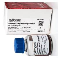
3)        Pipette to resuspend the beads completely in the sample lysate. Incubate with continuous mixing (rotating or roller mixer) for 3-5 min. at room temperature to allow the polyA tail of the mRNA to hybridize to the oligo(dT)25 on the beads. Increase the incubation time if the solution is viscous.

4)        Place the vial on the magnet for 2 min and remove the supernatant. If the solution is noticeably viscous, increase the time to approx. 10 min.

5)        Wash the beads/mRNA complex two times with the appropriate volume of Washing Buffer A at room temperature. Use the magnet to separate the beads from the solution between each washing step.

6)        Wash the beads/mRNA complex once with the appropriate volume of Washing Buffer B at room temperature Use the magnet to separate the beads from the solution.

7)        If the isolated mRNA is to be used in enzymatic downstream applications (e.g. RT-PCR), one extra wash in Washing Buffer B is recommended. This should be followed by a final wash in the enzymatic buffer to be used (e.g. RT-PCR buffer without the enzyme or primers).

Note:   Perform cDNA synthesis as recommended by the manufacturer of the reverse transcriptase. When using a thermostable reverse transcriptase and the bead-bound oligo (dT) as primer for first strand cDNA synthesis, an initial incubation at 50°C for 5 minutes is necessary before proceeding at the recommended temperature.

8)        If elution of mRNA from the beads is desired, add an appropriate volume of 10 mM Tris-HCl (Elution Buffer) and incubate at 65-80°C for 2 min. Immediately place the tube on the magnet, transfer the supernatant containing the mRNA to a new RNase free tube and place this tube on ice.

提问
扫一扫
丁香实验小程序二维码
实验小助手
丁香实验公众号二维码
扫码领资料
反馈
TOP
打开小程序