Construction and Hyperspectral Imaging of Quantum Dot Lysate Arrays
互联网
279
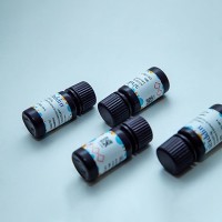
The emerging field of proteomic molecular profiling will be driven by new technologies that can measure dozens to hundreds of proteins from a small sample input from a patient’s biopsy. Lysate arrays, or reverse-phase protein microarrays, provide a platform for complex mixtures of proteins extracted from cells and tissues to be directly immobilized onto a solid support (such as a biochip with protein binding capacity) in diminutive volumes (picoliter-to-nanoliter). The proteins are spotted using precision robotics and then quantitatively assayed using primary antibodies; important posttranslational modifications, such as phosphorylations that are important for protein activation, may also be assayed to provide an estimate of the regulation of cellular signaling. Until recently, chromogenic signals and fluorescence (using organic fluorophores) detection were two strategies relied upon for signal detection. Emerging regents such as quantum dots (Qdot� nanocrystals; QD) are now employed for improved performance. QD embody a more versatile detection system because the robust signals may be time averaged and the narrow spectral emissions enable many protein targets to be quantified within the same lysate spot. Previously, we found that commercially available pegylated, streptavidin-conjugated QD were effective detection agents, with low-background affinities to spurious components within heterogeneous protein mixtures. Hyperspectral imaging allows the simultaneous detection of the different colored QD reagents within a single lysate spot. Here, we described the construction and imaging of QD lysate arrays. This technology is an emerging, enabling tool within the exciting, clinically oriented field of clinical tissue proteomics.