ELISPOT (Enzyme-linked ImmunoSPOT) 实验方法步骤
互联网
Introduction
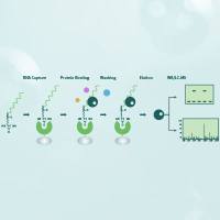
Enzyme-linked immunosorbent spot (ELISPOT) assays were originally developed to enumerate B cells secreting antigen-specific antibodies, but subsequently the assay has been adapted for identification and enumeration of cytokine-producing cells at the single cell level. The method employs the sandwich assay approach of the enzyme-linked immunosorbent assay (ELISA), with some variations. The ELISPOT capture antibody is coated aseptically onto a polyvinylidene difluoride (PVDF)-backed microwell plate. The plate is blocked with serum proteins, cells of interest are plated out at varying densities, along with antigen or mitogen, and plates are incubated at 37°C. Cytokine secreted by activated cells is captured locally by the coated antibody on the high surface area PVDF membrane. The wells are washed to remove cells, debris, and media components. A second antibody (biotinylated) reactive with a distinct epitope of the target cytokine is employed to detect the captured cytokine. The detected cytokine is then visualized using avidin-HRP, and a precipitating substrate (e.g., AEC). The colored end product (spot) represents an individual cytokine-producing cell. The spots can be counted manually (e.g., with a dissecting microscope) or using an automated reader to capture the microwell images and to analyze spot number and size.
The ELISPOT assay enables analysis of activated or responding cells at the single cell level. By virtue of exquisite sensitivity of the ELISPOT assay, frequency analysis of rare cell populations (e.g., antigen-specific responses) which were not possible by bulk assay methods are now possible. Limits of detection are below 1/100,000 rendering the assay uniquely useful for monitoring antigen-specific responses, applicable to a wide range of areas of immunology research, including cancer, transplantation, infectious disease, and vaccine development.
 |
 |
 |
 |
|
|
Human IL-17 ELISPOT: Left: Human PBMCs cultured for 24 hrs (no mitogen). Right: Human PBMCs activated with PMA/Ionomycin for 24 hrs |
Human Granzyme B ELISPOT: Left: Human PBMCs without mitogen cultured for 24 hrs. Right: Human PBMCs activated with PMA/Ionomycin for 24 hrs. |