Generation, Maintenance, and Differentiation of Human iPS Cells from Cord Blood
互联网
831
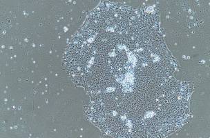
This chapter describes a robust method for the generation of iPS cells from non-cultured human cord blood cells. We describe the preparation of the CD34+ fraction from cord blood mononuclear cells, the protocols to determine the pluripotency of the reprogrammed cells, the culture conditions for serial passages of iPS cells, and the protocol for cryopreservation of established iPS cells. As the efficiency of gene transfer to suspension cells is relatively low compared with adherent cells such as fibroblasts or mesenchymal stem cells, improvements in the efficiency of gene transfer to cord blood cells is a key issue in using them to generate iPS cells. Here, iPS cells are generated by introducing Yamanaka’s four factors (Oct3/4, Sox2, Klf4, and c-Myc) with retroviruses, and, in addition, a short-hairpin RNA (shRNA) sequence with lentivirus that increases the rate of CD34+ cord blood cell proliferation. Enhanced green fluorescent protein (GFP) is also introduced to monitor the infection efficiency and the silence of exogenous genes. Infected cells are cultured in hematopoietic medium (X-VIVO 10 containing 50 ng/mL IL-6, 50 ng/mL soluble IL-6 R, 50 ng/mL SCF, 10 ng/mL TPO, and 20 ng/mL Flt-3-ligand) for 5 days after viral infection. Then, cells are transferred onto SNL76/7 feeder cells in human embryonic stem cell medium (DMEM/F-12 containing 20% KSR, 2 mM l -glutamine, 1% NEAA, 0.1 mM 2-ME, and 4 ng/mL bFGF). The ES cell-like colonies emerge in the following 4 weeks and are picked up to check their capacity for serial passage. The picked up colonies are stained to detect ALP activities and pluripotency-related molecules such as SSEA-4, TRA-1-60, and TRA-1-81 by immunochemistry. Further induction of endogenous pluripotency-related genes and silence of exogenous genes are determined by RT-PCR. The silencing of the introduced p53 knockdown lentiviral construct is checked by QRT-PCR. Finally, the differentiation potential of cells from ES cell-like colonies is determined by in vitro culture and teratoma formation assay with SCID mice. We explain each protocol in a practical way, focusing on problems we have encountered as stated in the Notes. We also utilize figures and photographs to facilitate comprehension of the protocols.