Linear amplification of limiting amounts of RNA for gene expression studies
互联网
Introduction
Expression profiling of cells is a valuable tool in understanding gene regulatory networks and in identifying genes necessary for normal developmental processes. In the haematopoietic system, cell surface markers are well defined allowing the isolation of cells from various developmental stages using flow cytometry. However gene expression studies of solid tissues are often hampered by the difficulty in obtaining a homogenous cell population. Often appropriate cell markers are not well defined for given cell stages therefore making gene expression studies difficult. In addition studies of rare cell populations mean that the amount of material obtained is insufficient for analysis. To overcome these problems we have employed a system where a given cell population is GFP-labeled and then sorted by flow cytometry in order to obtain a pure homogenous population. RNA was then prepared from this material and amplified by a linear amplification method allowing large scale microarray gene expression analysis. This approach was successfully applied to gene expression studies of the midbrain-hindbrain organizer region of the mouse but can be applied to many other systems to isolate cell populations.
Using the mouse as a model organism, our group investigated the role of the paired domain transcription factor Pax2 in brain development (Bouchard et al ., 2005). Pax2 is required for the formation of the isthmic organizer (IsO) at the midbrain-hindbrain boundary, where it initiates expression of the IsO signal Fgf8. To gain further insight into the role of Pax2 in mid-hindbrain patterning, our group searched for novel Pax2-regulated genes by cDNA microarray analysis of FACS-sorted GFP+ mid-hindbrain cells from wild-type and Pax2-/- E8.5 embryos carrying a Pax2GFP BAC transgene (Figure 1). Our sorting protocol yielded 5,000 to 10,000 GFP+ cells per embryo which was insufficient to generate enough RNA to hybridize to a microarray. Therefore RNA from the sorted cells was subjected to a linear amplification as described (Hoffmann et al ., 2003) with some modifications. Following this, the RNA was used in a microarray screen as described (Cheung et al ., 1999). Our unbiased chip approach identified the En2 , Brn1 , (Pou3f3 ), Sef , Tapp1 and non-coding Ncrms genes as genetic Pax2 targets that are totally dependent on Pax2 function for their expression in the mid-hindbrain region. Note that genes which showed only a 2 fold regulation on the microarray were confirmed as Pax2 target genes by in-situ hybridisation. This proves that the amplification protocol is linear and that genes with low induction ratios are indeed Pax2-regulated target genes (Figure 2).
Back to top
Procedure
Preparation of total RNA from limiting ex vivo sorted material
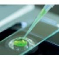
Sorting
Cells were sorted into 1.5ml microcentrifuge tubes containing 150µL FACS buffer. Before sorting, the inner walls of the tube were coated with buffer to prevent cells from sticking to the dry surface. After sorting, a small percentage of the sorted cells were used for reanalysis by FACS to verify the purity. If no reanalysis is necessary, the cells can be sorted directly into TRI reagent (Sigma).
RNA preparation
- Centrifuge cells at 13,000rpm for 30 seconds;
- Remove the supernatant leaving about 20µl remaining in the tube;
- Vortex the tube briefly to bring the cells into suspension again, and while vortexing, add 500µl TRIreagent;
- Leave the tube at room temperature for 5 minutes, and then add 5mg of carrier tRNA. Add 100µl of CHCI3 and centrifuge at 13,000rpm for 10 minutes;
- Transfer the upper phase into a new 1.5ml tube, and add 700ml of isopropanol;
- Precipitate the RNA by either putting the tube at -20°C o/n or at -80°C for 1 hour followed by centrifugation at 13.000rpm for 30 minutes;
- Wash the pellet with 75% EtOH (prepared with DEPC-H2 O) and dissolve in DEPC-H2 O (usually RNA from 50,000-100,000 cells are resuspended in 9ml).
Linear amplification of total RNA
Preparation of ds cDNA from total RNA
First strand synthesis (in RNAse-free PCR tubes)
- Dissolve RNA from at least 5,000 cells (ideally 50,000-100,000 cells) in 9ml DEPC-H2 O;
- Add 1µl PAGE-purified oligo-dT-T7-primer (500ng/µl), incubate the mix at 70°C for 10 minutes, snap-cool on ice;
- Prepare the following reverse transcription mix: 4µl 5x First strand cDNA buffer, 2µl 0.1M DTT, 1µl 10mM dNTP mix, 1µl RNAsin, 2µl Superscript II RT;
- Add 10µl mix to the RNA/primer sample and incubate at 42°C for 1 hour.
Second strand synthesis (in RNAse-free PCR tubes)
- Prepare the following mix: 86µl DEPC-H2 O, 30µl 5x second strand buffer, 3µl 10mM dNTP mix, 8µl DNA Polymerase I, 1µl E. coli DNA Ligase, 2µl RNAse H;
- Add 130µl mix to the first strand cDNA (total volume 150µl), tap the tube gently to mix;
- Spin briefly and incubate at 16°C for 2 hours;
- Add 3µl of T4 DNA polymerase;
- Incubate at 16°C for 5 minutes, put at 70°C for 10 minutes to stop the reaction.
- Extract the sample with 150µl Phenol/Chloroform;
- Add the upper aqueous phase from the extraction onto a Microcon-YM50 column already preloaded with 200µl DEPC-H2 O;
- Re-extract the organic phase with 150µl water;
- Load the upper aqueous phase from the second extraction onto the column;
- Centrifuge for 8 minutes at 9,000rpm, discard flow-through;
- Add 500µl DEPC-H2 O onto the column;
- Spin at 9,000rpm for 8 minutes, discard flow-through;
- Place the column inverted into a new tube, centrifuge at 2500rpm for 5 minutes tor elute;
- Bring the volume of the eluate to 8µl with DEPC-H2 O, in case of a bigger volume, speedvac to 8µl.
In vitro transcription (IVT) 1 (in RNAse-free PCR tubes)
- Add the IVT mix consisting of 4µl 5x IVT reaction buffer, 6µl 25mM rNTP mix and 2µl T7 enzyme mix to the ds cDNA (room temperature);
- Incubate at 37°C for 4 hours.
Clean-up (in RNAse-free microcentrifuge tubes)
- Add 130µl DEPC-H2 O to bring the total volume to 150µl;
- Purify aRNA using a Qiagen RNeasy kit according to the manufacturer’s protocol;
- Concentrate aRNA to 20µl in a speedvac.
Preparation of ds cDNA from aRNA
First strand synthesis (in RNAse-free PCR tubes)
- Use only half of the aRNA, i.e. 10µl, for further steps;
- Add 1,5µl of pd(N)9 (1µg/µl, dissolved in 10mM TRIS, pH 7.5) to the aRNA;
- Incubate the mix at 70°C for 10 minutes, snap-cool on ice.
Reverse transcription
- Prepare the following mix: 4µl 5x First strand cDNA buffer, 2µl 0.1M DTT, 1µl 10mM dNTP mix, 1µl RNAsin, 1µl Superscript II RT;
- Add 9µl mix to the aRNA/primer sample and incubate at 42°C for 1 hour;
- Add 3µl of RNAseH (removal of DNA/RNA hybrids);
- Incubate for 30 minutes at 37°C.
Second strand synthesis (in RNAse-free PCR tubes)
- Add 1.5µl PAGE-purified oligo-dT-T7-primer (500ng/µl);
- Incubate at 95°C for 5 minutes, snap-cool on ice, incubate at 42°C for 10 minutes;
- Add the following mix: 92µl DEPC-H2 O, 15µl 10x Eco-Pol buffer, 4µl 10mM dNTP mix, 8µl Klenow Enzyme and 8µl T4 DNA Polymerase;
- Incubate at 16°C for 2 hours.
Clean-up of double-stranded cDNA (in RNAse-free microcentrifuge tubes)
- Extract the sample with 150µl Phenol/Chloroform;
- Add the upper aqueous phase from the extraction onto a Microcon-YM50 column already preloaded with 200µl DEPC-H2 O;
- Re-extract the organic phase with 150µl water;
- Load the upper aqueous phase from the second extraction onto the column;
- Centrifuge for 8 minutes at 9,000rpm, discard flow-through;
- Add 500µl DEPC-H2 O onto the column;
- Spin at 9,000rpm for 8 minutes, discard flow-through;
- Place the column inverted into a new tube, centrifuged at 2500rpm for 5 minutes for elution;
- Bring the volume of the eluate to 8µl by adding DEPC-H2 O. If the eluate is more than 8µl then speedvac the sample to 8µl.
In vitro transcription (IVT) 2
- Add the IVT mix consisting of 4µl 5x IVT reaction buffer, 6µl 25mM rNTP mix and 2µl T7 enzyme mix to the ds cDNA (room temperature);
- Incubate at 37°C for 4 hours.
Clean-up (in RNAse-free microcentrifuge tubes)
- Add 130µl DEPC-H2 O to bring the total volume to 150µl;
- Purify aRNA using a Qiagen RNeasy kit according to the manufacturer’s protocol (see comment 1);
- Measure aRNA concentration on an Agilent Bioanalyzer 2001 according to the manufacturer's protocol.
The aRNA now can be used for microarray hybridizations. Follow standard microarray protocols for labeling and hybridizations. After 2 rounds of IVT, the amount of aRNA typically varies between 60-130µg when starting with 50,000 sorted cells. This is enough for at least 20 microarray hybridisations.
[1] [2] 下一页