Monitoring of the Dynamics of Epithelial Dome Formation Using a Novel Culture Chamber for Long-Term Continuous Live-Cell Imaging
互联网
392
Epithelial tissue guarantees proper performance of many organs, e.g., the kidneys, the gastrointestinal organs, and endocrine glands. Epithelial layers are responsible for the formation and maintenance of separate compartments with distinct solute composition. This is achieved by epithelial layers forming a barrier between the two compartments and concomitantly allowing site-directed transepithelial transport, uptake or secretion of electrolytes, energy substrates, proteins, and other solutes.
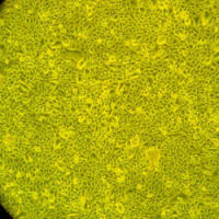
Research on epithelial tissue functions has highly profited from the establishment of tissue culture technologies allowing to cultivate primary epithelial cells or established epithelial cell lines. A property of transporting epithelia cultured in vitro that has long been noted is the formation of the so-called domes on solid growth supports, which represent fluid filled blisters between the solid growth surface and the cell layer. Formation of domes is regarded as a sign of active transport processes and an intact epithelial barrier function due to functional tight junctional cell–cell contacts. A novel methodology for long-term live-cell light microscopy is described in the present article, which allows the monitoring of the dynamic nature of structures, such as epithelial domes over days to weeks of tissue culture (“under the microscope”).