酶法、热修复、微波法修复抗原
互联网
If no antigen retrieval step is stated on the antibody data sheet, start off by trying the heat mediated pressure cooker method.
Frozen tissue does not need an antigen retrieval step. Once mounted on APES coated slides, they are best kept at -80°C until needed. When required, leave to warm at room temperature for 5 minutes, then acetone fix for 5 minutes followed by a PBS or TBS rinse. Afterwards, continue with the immunohistochemical staining protocol.
Enzymatic antigen retrieval
Tissue sections are best mounted on APES (amino-propyl-tri-ethoxy-silane) coated slides. Slides should be placed in a rack for this procedure.
Materials
Water baths
2 troughs
pH meter
Magnetic stirrer
Thermometer
Reagents
Alpha-Chymotrypsin (type II from Bovine pancreas) 0.1 g
Calcium Chloride 0.1 g
Ultra-pure water 100.0 ml
0.5% sodium hydroxide solution
0.5% hydrochloric acid solution
Xylene
Industrial methylated spirits (IMS) or methanol
Method
Set water bath to read 37°C. Add the required amount of ultra pure water into each trough then place the troughs into the water bath (See note i). Allow the ultra pure water warm to 37°C
Dewax and re-hydrate paraffin sections by placing them in 3 changes of xylene for 3 minutes each, followed by 3 changes of IMS or methanol for 3 minutes each, followed by cold running tap water for 3 minutes. At no time from this point onwards should the slides be allowed to dry out! Place slides in one trough of ultra pure water at 37°C to warm (See note ii).
Remove the other trough and into this dissolve the calcium chloride and chymotrypsin using a magnetic stirrer (See note iii). Once dissolved, pH to 7.8 using the 0.5% sodium hydroxide and hydrochloric acid solutions. Return the trough to the water bath and allow this enzyme solution to re-heat to 37°C (See note iv).
Transfer the warmed slides into the enzyme solution for a suggested 20 minutes (See note v) then remove the slides and place them into cold running tap water for 3 minutes (See note vi).
Continue with immunohistochemical staining protocol.
Notes
i) Use a sufficient volume of ultra pure water in order to cover the slides. Adjust the reagent quantities appropriately.
ii) Placing cold slides into the enzyme solution will lower the temperature of the solution, therefore reducing enzyme activity leading to the antigens being under-retrieved. Once the slides have been dewaxed and re-hydrated, drying out will cause non-specific antibody binding and therefore high background staining.
iii) Chymotrypsin can be very allergenic. Use a face mask and extraction cabinet for weighing out.
iv) Prepare the chymotrypsin solution as quickly as possible to avoid impairing the activity of the enzyme. Allow this solution to return to 37°C before introducing the slides.
v) 20 minutes is only suggested as a starting point incubation time. Less than 20 minutes may leave the antigens under retrieved, leading to weak staining. More than 20 minutes may leave them over retrieved, leading to non-specific background staining and also increasing the chances of sections dissociating from the slides. A control experiment is recommended beforehand, where slides of the same tissue section are incubated in the enzyme solution for 10, 15, 20, 25, and 30 minutes before being immunohistochemically stained to evaluate optimum antigen retrieval time for the particular antibody being used.
vi) Tap water stops the enzymatic process by washing action.
Heat mediated antigen retrieval
Pressure Cooker Method:
Tissue sections are best mounted on APES (amino-propyl-tri-ethoxy-silane) coated slides. Slides should be placed in a metal rack for this procedure.
Materials
Domestic stainless steel pressure cooker
Hot plate
pH meter
Magnetic stirrer
2 litre beaker or conical flask
Reagents
Tri-Sodium Citrate 5.88 g
0.2M Hydrochloric acid solution 44.0 ml
Ultra-pure water 1956.0 ml
0.5% sodium hydroxide solution
0.5% hydrochloric acid solution
Xylene
Industrial methylated spirits (IMS) or methanol
Method
Add the tri-sodium citrate, hydrochloric acid and ultra-pure water together in a 2 litre beaker/conical flask. Use a magnetic stirrer to ensure that all reagents are properly dissolved.
Adjust to pH 6.0 with 0.5% sodium hydroxide and hydrochloric acid solutions. Add this solution to the pressure cooker. Place the pressure cooker on the hotplate and turn it on full power. Do not secure the lid of the pressure cooker at this point, simply rest it on top.
While waiting for the pressure cooker to come to the boil, dewax and re-hydrate the paraffin sections by placing them in 3 changes of xylene for 3 minutes each, followed by 3 changes of IMS or methanol for 3 minutes each, followed by cold running tap water. Keep them in the tap water until the pressure cooker comes to the boil. At no time from this point onwards should the slides be allowed to dry out! (See note i.)
Once boiling, transfer the slides from the tap water to the pressure cooker. CARE WITH HOT SOLUTION - USE FORCEPS! Secure the pressure cooker lid as in the manufacturers instructions.
As soon as the cooker has reached full pressure (see the manufacturers instructions), time 3 minutes. (See note ii).
When 3 minutes has elapsed, turn off the hotplate and place the pressure cooker in an empty sink. Activate the pressure release valve (see the manufacturers instructions) and run cold water over the cooker. Once de-pressurised, open the lid and run cold water into the cooker for 10 minutes. CARE WITH HOT SOLUTION! (See note iii).
Continue with immunohistochemical staining protocol.
Notes
i) Once the slides have been dewaxed and re-hydrated, drying out will cause non-specific antibody binding and therefore high background staining.
ii) 3 minutes is only suggested as a starting point antigen retrieval time. Less than 3 minutes may leave the antigens under retrieved, leading to weak staining. More than 3 minutes may leave them over retrieved, leading to non-specific background staining and also increasing the chances of sections dissociating from the slides. A control experiment is recommended beforehand, where slides of the same tissue section are retrieved for 1, 2, 3, 4 and 5 minutes before being immunohistochemically stained to evaluate optimum antigen retrieval time for the particular antibody being used.
iii) This is to make the slides cool enough to handle and to allow the antigenic site to re-form after being exposed to such high temperature.
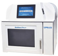
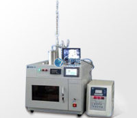
Microwave Method
The use of a domestic microwave is inadvisable. Hot and cold spots are common, leading to uneven antigen retrieval. Antigen retrieval times are usually longer, due to the absence of a pressurised environment, nearly always leading to section dissociation.
A scientific microwave is much more appropriate. Most brands have on-board pressurised vessels and can keep the temperature at a constant 98°C to avoid section dissociation. The only drawback is the expense of purchasing one!
Tissue sections are best mounted on APES (amino-propyl-tri-ethoxy-silane) coated slides. Slides should be placed in a plastic rack for this procedure.
Materials
Domestic (850W) or scientific microwave
pH meter
Magnetic stirrer
1 litre beaker or conical flask
Microwaveable vessel, either inbuilt or to hold approximately 400-500 ml
Reagents
Tri-Sodium Citrate 2.94 g
0.2M Hydrochloric acid solution 22.0 ml
Ultra-pure water 978.0 ml
0.5% sodium hydroxide solution
0.5% hydrochloric acid solution
Xylene
Industrial methylated spirits (IMS) or methanol
Method
Dewax and re-hydrate the paraffin sections by placing them in 3 changes of xylene for 3 minutes each, followed by 3 changes of IMS or methanol for 3 minutes each, followed by cold running tap water. Keep them in the tap water until the microwave antigen retrieval solution has been prepared. At no time from this point onwards should the slides be allowed to dry out! (See note i)
Add the tri-sodium citrate, hydrochloric acid and ultra-pure water together in a 1 litre beaker/conical flask. Use a magnetic stirrer to ensure that all reagents are properly dissolved. Adjust to pH 6.0 with 0.5% sodium hydroxide and hydrochloric acid solutions. Add this solution to the microwaveable vessel (See note ii).
Remove the slides from the tap water and place them in the microwaveable vessel. Place the vessel inside the microwave. If domestic, set to full power and wait until the solution comes to the boil. Time 15 minutes from this point. If scientific, program so that antigens are retrieved for 15 minutes once the temperature as reached 98°C. (See note iii).
When 15 minutes has elapsed, remove the vessel and run cold tap water into it for 10 minutes. CARE WITH HOT SOLUTION! (See note iv).
Continue with immunohistochemical staining protocol.
Notes
i) Once the slides have been dewaxed and re-hydrated, drying out will cause non-specific antibody binding and therefore high background staining.
ii) Use a sufficient volume of antigen retrieval solution in order to cover the slides. This should be by at least a few centimetres if using a non-sealed vessel so to allow for evaporation during the boil.
iii) 15 minutes is only a suggested antigen retrieval time. Less than 15 minutes may leave the antigens under retrieved, leading to weak staining. More than 15 minutes may leave them over retrieved, leading to non-specific background staining and also increasing the chances of sections dissociating from the slides. A control experiment is recommended beforehand, where slides of the same tissue section are retrieved for 5, 10, 15, 20, 25 and 30 minutes before being immunohistochemically stained to evaluate optimum antigen retrieval time for the particular antibody being used.
iv) This is to make the slides cool enough to handle and to allow the antigenic site to re-form after being exposed to such high temperature.
上一篇:免疫组化石蜡包埋组织抗原修复 下一篇:高温处理+柠檬酸钠或EDTA修复石蜡包埋样品抗原