Reflection Contrast Microscopy: The Bridge Between Light and Electron Microscopy
互联网
674
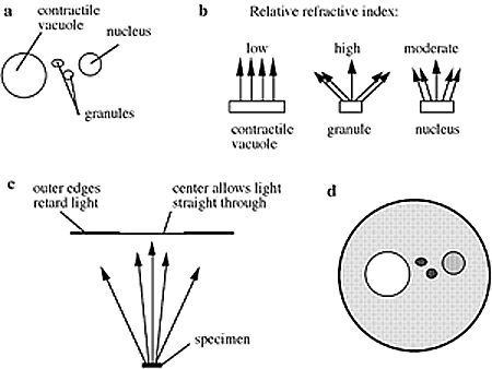
Reflection contrast microscopy (RCM) is a light microscopic method to image cells at high definition and enhanced sensitivity compared to conventional bright-field microscopy. RCM images have very high contrast, which makes them easily applicable for digital image analysis. Because ultrathin sections are mostly used in this method, RCM also functions by bridging light with electron microscopy: the combination of ultrastructural with histochemical studies. RCM can also replace electron microscopy for rapid and simple screening of large quantities of samples for immunocytochemical staining. Special attention is paid to small biological objects, which have to be processed for RCM. If you encounter the limits of brightfield microscopy, in resolution, sensitivity or handling of the specimen, RCM will be a feasible option.
Reflection contrast microscopy methods use only slightly adjusted electron microscopy methods for specimen preparation. Therefore, many familiar techniques for ultrathin specimen preparation can be applied. It is essential that only refractive index differences exist in those areas that are of interest and that the further specimen is as optically homogenic as possible, with a refractive index as close to that of glass as possible. Therefore, plastic embedding is recommended.