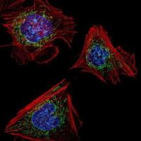
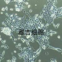
MG63细胞
互联网
摘 要
一、人成骨肉瘤MG-63细胞株和成人成骨细胞的培养与分化特性的观察
目的:观察人成骨肉瘤MG-63细胞株和正常成人成骨细胞分化的特性。方法:在细胞培养的不同时间,用α-磷酸奈酚法测定碱性磷酸酶(ALP)活性;放射免疫法测定骨钙素(BGP)含量;半定量逆转录聚合酶链反应(RT-PCR)测I型胶原、MMP-1、TIMP-1基因mRNA表达;Van GieSon氏苦味酸酸性复红染色法染色细胞I型胶原。结果:人成骨肉瘤MG-63细胞株接种培养第7天后I型胶原基因表达较高。
MMP-1表达量随时间推移逐渐增加,至第24天达到高峰。第1~9天TIMP-1 表达量逐渐增加,其后基本恒定。0~12天ALP活性逐渐增高,致12天达最高,其后逐渐下降。18天后,细胞有许多大小不等的结节形成,I型胶原结节染色较无结节处深。正常成人成骨细胞(hOB)接种培养后0~6天细胞逐渐汇片。0~12天ALP活性逐渐增高,致12天达最高,其后逐渐下降。第6天BGP表达最高,其后逐渐下降。结论:分离培养的hOB具有成骨细胞的特性;人成骨肉瘤MG-63细胞株为具有人成骨细胞表型特征的成骨细胞模型。人成骨肉瘤MG-63细胞株和hOB的生长分为细胞增殖、骨基质成熟、骨基质矿化阶段,且I型胶原、BGP、MMP-1、TIMP-1基因表达及ALP活性呈时间特异性。
二、人成骨肉瘤MG-63细胞株和成人成骨细胞基质金属蛋白酶及其抑制因子的表达
目的:观察人成骨肉瘤MG-63细胞株和正常成人成骨细胞(hOB)基质金属蛋白酶(MMPs)和基质金属蛋白酶抑制因子(TIMPs)表达的类型。方法:MMPs和TIMPs mRNA表达用RT-PCR检测。结果:人成骨肉瘤MG-63细胞株和hOB均表达MMP-1、MMP-2、MMP-7、MMP-10、MMP-11、MMP-14、MMP-15、MMP-16、MMP-17、TIMP-1、TIMP-2、TIMP-3。人成骨肉瘤MG-63细胞株可表达MMP-12,hOB则无。结论:人成骨肉瘤MG-63细胞株和hOB表达的MMPs和TIMPs类型基本相似,人成骨肉瘤MG-63细胞株可以作为具有人成骨细胞表型特征的成骨细胞模型来研究MMPs和TIMPs的表达与调控。
三、雌二醇对人成骨肉瘤MG-63细胞株和成人成骨细胞基质金属蛋白酶及其抑制因子的作用
目的:探讨雌二醇(17β-estradiol,E2)对人成骨肉瘤MG-63细胞株和正常成人成骨细胞(hOB)基质金属蛋白酶(MMPs)及其抑制因子(TIMPs)的作用和雌激素缺乏致骨质疏松的发病机制。方法:MMPs和TIMPs蛋白质表达用ELISA和Western Blot检测, mRNA用RT-PCR检测。MMPs活性用明胶酶谱法检测。
结果:首次发现E2抑制人成骨肉瘤MG-63细胞株MMP-1 mRNA和蛋白质表达,并且呈剂量依赖关系;10-8M E2抑制hOB MMP-1蛋白质表达;E2对人成骨肉瘤MG-63细胞株MMP-2及TIMP-1 mRNA及蛋白质表达无影响;E2对hOB MMP-2及TIMP-1 蛋白质表达无影响;E2对人成骨肉瘤MG-63细胞株和hOB MMP-2活性亦无影响。
结论:雌激素不足可导致其对人成骨样细胞MMP-1的抑制作用减弱,促进骨吸收和骨基质分解,此可能为雌激素缺乏致骨质疏松的重要发病机制。
四、雌二醇对人成骨肉瘤MG-63细胞株和成人成骨细胞膜型 基质金属蛋白酶-1的影响
目的:膜型基质金属蛋白酶-1(membrane type 1-matrix metalloproteinase, MT1-MMP)是最近发现的一种可由多种细胞表达的基质金属蛋白酶。为探讨雌二醇(E2)对人成骨肉瘤MG-63细胞株和正常成人成骨细胞(hOB)膜型基质金属蛋白酶(MT1-MMP)的作用和绝经后骨质疏松症(OP)的作用机制。
方法:Northern杂交、Western杂交和激光共聚焦显微系统免疫荧光法分别检测MT1-MMP mRNA和蛋白质表达。MMP-2活性用明胶酶谱和酶联免疫吸附测定检测。结果 首次观察到E2促进MG-63细胞MT1-MMPmRNA表达,并呈剂量依赖关系。E2促进MG-63细胞MT1-MMP蛋白质表达,并呈剂量依赖关系;E2干预24~48小时促进MG-63细胞MT1-MMP蛋白质表达;激光共聚焦显微系统免疫荧光法证实E2促进MT1-MMP蛋白质表达增强,MT1-MMP蛋白质表达于细胞膜和细胞质中。E2对MG-63细胞MMP-2活性无影响。
结论:E2可促进MG-63细胞MT1-MMP表达。雌激素不足可减少成骨样细胞MT1-MMP表达,此可能为绝经后OP的骨吸收增强机理之一。
Abstract
Part One Oberservation of the Differentiation of Human Osteosarcoma Cell Line MG-63 and Human Osteoblast in Culture
Objective: To observe the differentiation of human osteosarcoma cell line MG-63 and normal human osteoblast(hOB) in culture. Methods: Alkaline phosphatase(ALP) activity was determined by using p-nitrophenyl phosphate as substrate. Bone Gla protein(BGP) was measured by radioimmunoassay.Type I collagen,MMP-1,TIMP-1 mRNA was examined using semiquantitive reverse transcription-polymerase chain reaction(RT-PCR) analysis.Human osteosarcoma cell lines MG-63 were stained by the Van GieSon procedure. Results: In human osteosarcoma cell line MG-63 culture Type I collagen mRNA expression reach a maximum level on days 7.MMP-1 mRNA was not expressed until the fifth day of culture and increased with time developing.TIMP-1 mRNA was nearly constant. ALP activity decreased by days 18 after gradually up-regulated during the period from 0-12 days and reached it peak by days 12.Van GieSon staining displayed nodule formation at 12 days and became more prominent by days 18 in MG-63 cells culture.Development of ALP activity in hOB culture was similar to human osteosarcoma cell line MG-63 culture,the levels of BGP increased from days 3-6 and then steadily decreased by days 18. Conclusion: Human osteosarcoma cell line MG-63 and hOB cultured by us have the osteoblast phenotype.The are three principle periods during the differentiation of human osteogenic sarcoma cell line MG-63 and hOB-proliferation,extracellular matrix maturation,and mineralization.Gene expression in human osteosarcoma cell line MG-63 and hOB culture diplay temporal sequence.
Part Two Expression of Metalloproteinases and Tissue Inhibitors of Metalloproteinases in Human Osteoblast-like cells
Objective: To observe the differentiation of human osteosarcoma cell line MG-63 and human osteoblast(hOB) in culture. Methods: Metalloproteinases(MMPs) and tissue inhibitors of metalloproteinases(TIMPs) mRNA was examined using semiquantitive RT-PCR analysis. Results:MMP-1,MMP-2,MMP-7,MMP-10,MMP-11,MMP-14,MMP-15,MMP-16,MMP-17,TIMP-1,TIMP-2 and TIMP-3 were expressed by both human osteosarcoma cell line MG-63 and hOB.MMP-12 was expressed by human osteosarcoma cell line MG-63,but was not measured in hOB. Conclusion: Human osteosarcoma cell line MG-63 could been used as models for the osteoblast phenotype to understand the expression and regulation of MMPs and TIMPs.
Part three Effects of 17β-Estradiol(E2) on the Expression of Matrix Metalloproteinases and Tissue Inhibitor of Matrix Metalloproteinases in Human Osteoblast-like Cells
Objective: To characterize the effects of E2 on the expression of matrix metalloproteinases (MMPs) and tissue inhibitor of matrix metalloproteinases (TIMPs) in human osteosarcoma cell line MG-63 and normal human osteoblast(hOB) in order to understand the mechanism of estrogen deficient osteoporosis. Methods: Reverse transcription polymerase chain reaction(RT-PCR) was used to analyse the expression of MMPs and TIMPs mRNA under conditions of MG-63 cells with or without treatment of E2. ELISA and Western Blot were used to show the actions of E2 on secretion of MMPs and TIMPs of MG-63 cells and hOB. Gelatin zymogram was done to examine the effects of E2 on activity of MMPs. Results:Treatment with increasing dose of E2 in MG-63 cells caused a dose-dependent decrease in expression of MMP-1 mRNA and protein. 10-8M E2 inhibit the secretion of MMP-1 in hOB culture. E2 seemed to have no influence on the expression of MMP-2 and TIMP-1 mRNA and protein in MG-63 cell and hOB culture,it also has no effect on the activity of MMP-2. Conclusion: Deficiency of E2 as seen in postmenopausal women might decrease the inhibiting effect on MMP-1 in human osteoblastic-like cells, which could lead to the increasing degradation of bone matrix and bone resorption followed by acceleration of bone loss. It probably is one of the key pathogenesis of estrogen deficient osteoporosis.
Part four Effects of 17β-Estradiol (E2) on the Expression of Membrane Type 1-Matrix Metalloproteinase (MT1-MMP) in Human Osteoblast-like Cells
Objective: To characterize the effects of E2 on the expression of MT1-MMP in human osteosarcoma cell line MG-63 in order to understand the mechanism of estrogen deficient osteoporosis. Methods Northern Blot, Western Blot and confocal immunohistochemistry were respectively used to show the actions of E2 on expression of MT1-MMP mRNA and protein of MG-63 cells. Gelatin zymogram and ELISA were done to examine the effects of E2 on activity of MMP-2. Results Treatment with increasing dose of E2 in MG-63 cells caused a dose-dependent enhancement in expression of MT1-MMP protein (P<0.05), furthermore confirmed by confocal immunohistochemistry. E2 seemed to have no influence on the activity of MMP-2 in MG-63 cells (P>0.05). Conclusion Deficiency of E2 as seen in postmenopausal women may inhibiting the expression of MT1-MMP in human osteoblast-like cells, leading to the acceleration of bone resorption. It probably is one of the key pathogenesis of estrogen deficient osteoporosis.


