线粒体凋亡的信号通路图
互联网
- 细胞凋亡专题
相关专题
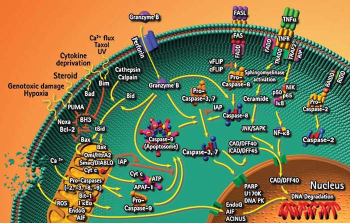
线粒体 凋亡途径是细胞凋亡的主要途径之一,是目前研究凋亡的热点.各种凋亡刺激信号通过BH3(Bcl-2 homology domain 3)-only蛋白引起Bax(Bcl-2-asslciated protein X)蛋白移位到线粒体外膜并多聚化,形成膜通道,刺激线粒体释放细胞色素C(Cyt C) 和Smac(second mitochondrial-derived activator of caspase),Cyt C通过Apaf-1因子的多聚化与胱天蛋白酶(caspases)-9形成凋亡小体,导致下游胱天蛋白酶的级联反应。
而凋亡蛋白抑制因子(IAP)和Smac通过抑制和促进胱天蛋白酶的级联反应来调控细胞凋亡。
The Bcl-2 family of proteins regulate apoptosis by controlling mitochondrial permeability and the release of cytochrome C. The anti-apoptotic proteins Bcl-2 and Bcl-xL reside in the outer mitochondrial wall and inhibit cytochrome C release. The pro-apoptotic Bcl-2 proteins Bad, Bid, Bax and Bim reside in the cytosol but translocate to mitochondria following death signaling, where they promote the release of cytochrome C. Bad translocates to mitochondria and forms a pro-apoptotic complex with Bcl-xL. This translocation is inhibited by survival factors that induce the phosphorylation of Bad, leading to its cytosolic sequestration. Cytosolic BID is cleaved by caspase 8 following signaling through Fas: its active fragment (tBid) translocates to mitochondria. Bax and Bim translocate to mitochondria in response to death stimuli, including survival factor withdrawal.p53, activated following DNA damage, induces the transcription of Bax. Released cytochrome C binds Apaf1 and forms an activation complex with caspase 9. Although the mechanism(s) regulating mitochondrial permeability and the release of cytochrome C during apoptosis are not fully understood, Bcl-xL, Bcl-2 and Bax apparently influence the voltage-dependent anion channel (VDAC), which can control cytochrome C release.