Immunohistochemistry on fixed, paraffin-embedded sections
互联网
Immunohistochemistry on fixed, paraffin-embedded sections.
Rationale:
Immunostaining on formalin-fixed, paraffin embedded sections has been revolutionized in 1991 by the discovery of heat-mediated retrieval (Antigen Retrieval, AR) of immunoreactivity (Ref.
1 , 2
). This method is now widely used and applies to the detection of the overwhelming majority of antigens, with few exceptions for which enzymatic retrieval is required (Ref.
3
). The method shown here uses a strong chelating agent, EDTA, (Ref.
4
) ,and has been found efficient for the majority of antigens investigated.
More references are linked
here .
In order to enhance adhesion, the usual recommendation is to "bake" the slides (30 min at 6undefinedC or overnight (ON) at 37°C.)
We found this procedure unnecessary if the slides are prepared correctly and when slides are not dried in vertical position or the water is dirty, or there are air bubbles, baking does not helps at all. Anyway, do not exceed 6undefinedC and/or 30 min. for drying. Do not use microwave oven for drying.
Deparaffinize the slides with two 5 min. incubations of clean xylene, followed by three washes with absolute ethanol. Then gradually bring to distilled water.
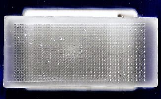
Place the sections in a radiotransparent slide holder (not metal; WVR/Baxter slide staining holder S7636. A good alternative are those plastic slide holder for shipping slides in the mail: ask your pathologist). <center> <span>click to enlarge the image</span> <br /> </center>
Immerse slides and holder in 1mM EDTA pH 7.5 (from a 100mM stock) in a beaker. Cover with a piece of Saran wrap in which you made holes. Put in a microwave oven and bring to a boil at max power (8 min for 800 ml). Let boil for 15 min at a reduced power (Power 3) so that the liquid continue to simmer. Cool at RT for 30 min to one hour. Transfer to TBS.
Alternatively a pressure cooker or an autoclave can be used, with optimal results.
You will perform all stainings in a humid chamber, as the one depicted below.
NOTE : The moist chamber (see above) must fulfill the following criteria: a) be absolutely flat, b) maintain moisture over 3 days without drying. c) be washable.Double indirect AP immunohistochemistry (e.g rabbit anti antigen X, goat anti-rabbit AP, rabbit anti-goat AP)
1- Wash twice in TBS 0.05M pH7.5, to which 0.01% Tween 20 has been added (TBS-T).
2- Briefly blot the slides without letting them dry and then apply 3% human or pig serum in PBS-BSA-NaN 3 as a blocking agent (health hazard!).
3- incubate with the blocking for 10 min. If one of your antibody is biotin-conjugated, you need at this point to do endogenous biotin blocking .
4- blot the slides without washing and apply the primary antibody (100µl), in a moist chamber, at RT for 1-18 hr.
5- Wash twice in TBS-T. 5 min / wash.
6- add the AP conjugated secondary antibody (50 to 100 µl) and incubate for 45 min. The secondary antibody should be absorbed against human serum; if not add 1% human serum before use. SBA Goat anti mouse AP or Goat anti Rabbit AP can be used 1:200 in TBS-BSA- NaN 3 .
7- Wash thrice in TBS-T.
[8 and 9 are optional]
8- add the AP conjugated tertiary antibody (50 to 100 µl) and incubate for 15 min. The tertiary antibody should be absorbed against human serum; if not add 1% human serum before use. SBA Rabbit anti Goat-AP or Goat anti Rabbit AP can be used 1:300 and 1:200 respectively in TBS-BSA NaN 3 .
9- Wash thrice in TBS-T.
10- add 50 ml of the developing solution (see below ). Protect from direct light.
11- after 5 min, check the staining in your positive and negative controls.
12- check the staining at 10-15 min interval.
13- when staining is complete (usually < 1 hr), wash thoroughly in tap water.
14- preferably postfix in formalin for 4-5 hrs before mounting in water soluble mounting medium (glycerol gelatin).
AP Developing solution:
For 50 ml developing solution add in order:
- 50 ml Tris-Hcl 0.1M pH 9.2 (exact!) (1:10 from a stock solution 1M)
- Levamisole 1mM (12 mg/50 ml) (Sigma L9756)
- 20 mg Naphtol As BI phosphate (Sigma N2250, the cheapo) (stock solution 40 mg/ml in NN-DM formamide, anhydrous, 0.5 ml aliquots kept at -20°C)
- 10 mg Fast Blue BB Diazonium (Sigma F3378) salt or Fast Red Diazonium salt
Filter with a 45µm filter.
Keep away from direct light, use within 5 min.
Indirect immunohistochemistry with avidin-biotin peroxidase(e.g rabbit anti antigen X, goat anti-rabbit biotin, avidin HRP)
1- Wash twice in TBS-T. 5 min / wash.
2- Briefly blot the slides without letting them dry and then apply egg white in PBS-BSA-NaN 3 as a blocking agent (one egg white in 100 ml PBS + NaN 3 ) (Miller RT et al, Appl Immunohistochemistry 5(1): 63-66, 1997). Incubate for 10 min. minimum. (Use the yolk as directed in www.cucinait.com ). Alternatively, use commercial blocking kits. Wash briefly once.
3- Apply 3% human or pig serum a s a blocking agent (health hazard!) for 10 min [after egg this is sometimes unnecessary]. Wash briefly.
4- Incubate with skim milk 5% in TBS-T for 10-30 min. (Miller RT et al, Appl. Immunohist & Molecular Morphology, 71(1): 63-65, 1999). This is the free avidin blocking step. Wash twice in TBS-T.
5- blot the slides without washing and apply the primary antibody, in a moist chamber, at RT for 1-18 hr.
6- Wash twice in TBS-T. 5 min / wash.
7- add the biotin-conjugated secondary antibody (50 to 100 µl) and incubate for 45 min. The secondary antibody should be absorbed against human serum; if not add 1% human serum before use. Most reagents are used at 1:100 or 1:200 dilution.
8- Wash twice in TBS-T.
8bis- block endogenous peroxidase by incubating in 0.1%NaN 3 and 0.3% H 2 O 2 for 30 min. Wash thrice.
9- add the HRP-conjugated avidin (50 to 100 µl, dilution 1:300 -500) (Dako P0364) and incubate for 20 min. Be careful not to dilute the avidin-HRP in biotin-containing or azide-containing medium.
10- Wash thrice in TBS-T.
11- add 50 ml of the developing solution (see below ). Protect from direct light.
12- after 5 min, check the staining in your positive and negative controls.
13- check the staining at 10-15 min interval.
14- when staining is complete (usually < 1 hr), wash thoroughly in tap water.
15- counterstain and mount in water soluble mounting medium (glycerol gelatin).
HRP Developing solution:
For 50 ml developing solution add in order:
- Aminoethylcarbazole (20 mg tablets, Sigma A-6926, dissolved in 2.5 ml NN-DM formamide)
- 50 ml acetate buffer pH 5.5 (52.5 ml of 0.1M acetic acid solution + 196.5 ml of a 0.1M Na acetate solution, bring to 500 ml)
- 25 µl H2O2 30%.
Filter with a 45µm filter (optional).
Keep away from direct light, use within 5 min. <center> <a name="HRP Developing solution"></a> <p> <a name="HRP Developing solution"> </a> </p> </center>
Double Immunohistochemistry 下一篇:In situ hybridization for Epstein Barr Virus-encoded RNA (EBER)

![Hep Par 1 Antibody / Hepatocyte Paraffin 1 [clone OCH1E5] (V2341)](https://img1.dxycdn.com/p/s14/2025/0214/113/8001790498709820981.jpg!wh200)


