Working Procedures in Microwave Histology
互联网
Working Procedures in Microwave Histology
by Vincent R. Klump, Jr., HT (ASCP), Histology Services, East Haven, CT
note: Mr. Klump was instrumental in the development of a standardized, consistent, reproducible protocol for microwave histoprocessing in the original Energy Beam Sciences H2500 Microwave Processor.
Microwave Tissue Processing
Tissue criteria: Maximum tissue size for this procedure: 3mm x 3mm x 3mm. Variations of this size can also be accommodated, but total tissue volume should not exceed 3mm cubed.
Examples: 3mm punch biopsies, core needle biopsies, core bone marrow biopsies, shave biopsies.
Tissue should be fixed in 10% NB Formalin for a minimum of four hours.
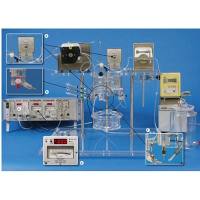
Equipment:
- Laboratory microwave equipped with a temperature probe accurate to +/- 1ºC
- Microwave-safe container holding a minimum of 250ml
- (optional) microwave-safe cassette rack supplied by Energy Beam Sciences
- 100% ethanol
- 100% isopropanol
- liquid paraffin
- boiling chips (preferably, common stones not bigger than 1cm x .5cm x .5cm)
Procedure:
- Rinse tissue for 5 minutes in running water
- dehydrate in 100% ethanol for 15 minutes at 65ºC in microwave
- clear in 100% isopropanol for 10 minutes at 74ºC in microwave
- infiltrate (3 steps) in liquid paraffin for 5 minutes at 65ºC in microwave with boiling chips, 5 minutes at 74º in microwave with boiling chips; fresh change of liquid paraffin for 5 minutes at 82º in microwave with boiling chips
- transfer cassettes to clean paraffin and embed as usual
Antigen Retrieval using Citrate Buffer
Slide criteria: Positively (+) charged or silane treated precleaned glass slides. Tissue placed centrally on slides. Slides baked for a minimum of 1 hour at 60ºC (preferably, baked overnight at 60ºC)
Examples:
Antigen retrieval may be utilized for almost all antibodies, enhancing staining and allowing for greater antibody dilution as well as shorter incubation times. Most antibodies which require enzyme treatment stain remarkably well utilizing antigen retrieval procedures. Remember to always run a known positive control with each antibody and a true negative with each case.
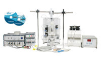
Equipment:
- Laboratory microwave with temperature control accurate to +/- 1ºC
- Microwave-safe staining dish
- Microwave-safe slide rack
- Distilled water
- Citric Acid
- Sodium Hydroxide
- pH meter
Procedure:
- Xylene, 2 changes, 10 minutes each
- 100% ethanol, two changes, 1 minute each
- 2% Hydrogen Peroxide Methanol quench for 10 minutes
- 100% ethanol, two changes, 1 minute each
- 95% ethanol, two changes, 1 minute each
- 70% ethanol, 1 minute
- Rinse in distilled water, 1 minute
Citrate buffer: 10mM Citrate Buffer (2.1g Citric Acid to 1L of distilled water, adjust pH to 6.0 with approximately 10mL of 2M NaOH). Immerse sections in citrate buffer for 10 minutes at 100ºC in microwave.
Note: Slides *musundefined cool to room temperature before proceeding to IHC steps. Rinse slides in distilled water, transfer to buffer, and go on to IHC procedure. A variety of staining techniques are suited to this procedure, including APAAP, PAP and ABC Complex/HRP.
Note: This procedure was adapted from Dako Antibody Data Sheet for KI-1
Special Stains Utilizing the Microwave PAS, PAS + DIASTASE, Fe++
We are all very familiar with silver stains in the microwave, but these three stains are histochemical stains which the microwave accelerates considerably.
PAS + DIASTASE Procedure:
PAS + DIASTASE done at room temperature takes two hours. PAS + DIASTASE aided by the microwave takes 50 minutes.
Equipment:
- Laboratory microwave with temperature control accurate to +/- 1ºC
- Microwave-safe Coplin jars
- .05% Periodic Acid
- Schiff's Reagent
- Hematoxylin, Gill III
- Diastase of malt, reagent grade, .5 gram/50ml distilled water (mix well, make fresh)
Procedure:
- Bake, deparaffinize and hydrate slides according to standard protocol
- Microwave slides for 15 minutes at 37ºC in diastase solution
- Rinse for 10 minutes in running water
- .05% Periodic Acid for 5 minutes
- Rinse for 1 minute in running water
- Microwave slides for 1 minute at 30ºC in Schiff's solution (be sure to either mix solution or use air bubble agitator to equalize temperature).
-
Leave slides for 5 minutes in this solution.
Note: This step *musundefined be done under a hood. - Wash in tap water at room temperature until a pink color develops (at least 1 minute).
- Counterstain in Gill III Hematoxylin for 1 minute
- Blue rinse in tap water; dehydrate, clear and mount
Note: This procedure was adapted from "The Microwave Cookbook for Microscopists", Boon and Kok, Coulomb Press Leiden, 1992
PAS Procedure:
PAS done at room temperature takes 45 minutes. PAS aided by the microwave takes 15 minutes.
Equipment:
- Laboratory microwave with temperature control accurate to +/- 1ºC
- Microwave-safe Coplin jars
- .05% Periodic Acid
- Schiff's Reagent
- Hematoxylin, Gill III
Procedure:
- Bake, deparaffinize and hydrate slides according to standard protocol .05% Periodic Acid for 5 minutes
- Rinse for 1 minute in running water
- Microwave slides for 1 minute at 30ºC in Schiff's solution (be sure to either mix solution or use air bubble agitator to equalize temperature).
-
Leave slides for 5 minutes in this solution.
Note: This step *musundefined be done under a hood.
- Wash in tap water at room temperature until a pink color develops (at least 1 minute).
- Counterstain in Gill III Hematoxylin for 1 minute
- Blue rinse in tap water; dehydrate, clear and mount
Note: This procedure was adapted from "The Microwave Cookbook for Microscopists", Boon and Kok, Coulomb Press Leiden, 1992
Fe++ Procedure:
Fe++ done at room temperature takes 30 minutes. Fe++ aided by the microwave takes less than 2 minutes.
Equipment:
- Laboratory microwave with temperature control accurate to +/- 1ºC
- Microwave-safe Coplin jars
- 2% Potassium Ferrocyanide
- 2% Hydrochloric Acid
- Nuclear Fast Red Solution
Procedure:
- Bake, deparaffinize and hydrate slides according to standard protocol
- Prepare working solution:
- 25ml of 2% Potassium Ferrocyanide, 25ml of 2% Hydrochloric Acid (prepare fresh before use)
- Place slides in microwave-safe Coplin jar with 50ml of working solution
- Microwave for 40 seconds at high power (do not use temperature probe in this step, as there is a potential for reaction between metal probe and working solution)
Note: All microwaves vary in power output; adjust microwave power level to acheive final temperature of 60ºC. Rinse well in distilled water. Counterstain in Nuclear Fast Red for 15 seconds at 60ºC using temperature probe Rinse in tap water, dehydrate, clear and mount
Note: This procedure was adapted from "The Microwave Cookbook for Microscopists", Boon and Kok, Coulomb Press Leiden, 1992
Microwave Stimulated Decalcification
Examples:
Bone biopsies are usually decalcified for a period ranging from one hour to days in a weak hydrochloric acid solution or in EDTA. With the aid of microwave exposure, the time is accelerated from one hour using conventional methods to as little as 10 minutes for bone marrow core biopsies using formic acid.
Equipment:
- Laboratory microwave equipped with a temperature probe accurate to +/- 1ºC
- Microwave-safe container holding a minimum of 250ml
- (Optional) Microwave cassette rack supplied by Energy Beam Sciences
- 5% Formic Acid Solution
Procedure:
- Keep bone biopsies as thin as possible
- Fix for standard times in 10% NB Formalin
- Place the bone biopsies in a microwave-safe container with at least 10x volume of 5% Formic Acid Solution to volume of tissue
- Microwave for 10 minutes at 55ºC
- Repeat this procedure until desired softness is achieved
- Rinse in running tap water for at least 10 minutes
- Proceed with either conventional or microwave processing
Note:
Treatment time depends on thickness and on density: solid bone and teeth take more time than spongy bone. Bone marrow biopsies require as little as 10 minutes exposure time.
Microwave Stimulated Fixation: Advantages and Disadvantages
Examples:
Standard fixation protocols in formalin take a minimum of four hours at room temperature. Fixation aided by microwave exposure and pre- and post-treatment in formalin take less than 30 minutes.
Equipment:
- Laboratory microwave equipped with a temperature probe accurate to +/- 1ºC
- Microwave-safe container holding a minimum of 500ml
- (Optional) Microwave cassette rack supplied by Energy Beam Sciences
- PBS 0.1M pH 7.4
- 10% NB Formalin
- 70% Ethanol
Procedure:
- Place tissue in 10% NB Formalin as a transport or collection medium
- Gross specimens no larger than 3mm in thickness
- Place in plastic cassettes
- Collect specimens in groups of 24 for fixation
- Immerse specimens in approximately 500ml PBS 0.1M pH 7.4 (cassette rack may be used, or groups of 20 specimens in cassettes in a suitable microwave-safe container)
- Microwave at 68ºC for 5 minutes
- Place tissue in 70% Ethanol
- Continue with conventional histoprocessing or microwave histoprocessing
Advantages:
10% NB Formalin can virtually be eliminated from the laboratory. Immunostaining is greatly enhanced in microwave-fixed tissue. Time is saved versus conventional 4 hour fixation .
Disadvantages:
Many laboratories are generally resistant to changes in established procedures. No control of time in formalin when specimens are received from outside the lab. Immunostaining results may vary due to changing times in formalin and increased cross-linking.
Conclusion:
Many factors must be considered when thinking about switching to microwave fixation of tissue rather than conventional fixation. First, the manner in which specimens are transported to your laboratory is the most critical factor in controlling pre-fixation time in formalin. Any reference laboratory or laboratory receiving specimens through the mail generally receive these specimens already fixed. These laboratories can consider removing formalin from the tissue processor and storing specimens in 70% Ethanol for a more chemically-friendly laboratory.
The laboratories which process samples biopsied at their own medical center or picked up by a local courier can best utilize microwave fixation. These labs can control times in formalin pre-fixation, which can lead to more consistent immuno results.
Another factor to be considered is the change in scheduling in the standard laboratory routine. Scheduling changes will affect everyone, from the courier to the clerical people involved in reporting.
I conclude that microwave fixation is not adaptable to every laboratory, but, in those situations where specimen transport can be regulated, it can be both cost-effective and more healthy for technicians and the environment. Keep in mind that any laboratory can utilize this time-saving procedure for stat biopsies, which can ultimately contribute to better patient care and a good option for fast specimen turn-around.
Slide Drying in the Microwave Example:
Slide drying in a conventional oven is done at temperatures between 60ºC and 80ºC, for times ranging from 20 to 60 minutes. Slide drying in a microwave takes from 2 to 5 minutes.
Equipment: Laboratory microwave with output power from 600-900 watts and equipped with a carousel. Microwave-safe slide racks holding 20-24 slides
Procedure:
- Cut standard paraffin sections from 4-6um and place on glass microscope slides
- Place slides in microwave-safe slide rack
- Drain water off of slides by gently tapping over paper towel or blotter
-
Place slides centrally on microwave carousel and microwave at high power for 2-5 minutes
Note: Times will vary with output power of microwave, and must be determined experimentally in each laboratory with its microwave
- Let slides briefly cool and continue with standard staining procedure
Note: When consequetive batches are dried in a microwave, the microwave cavity temperature will get gradually higher. When not drying slides, keep cavity door open to allow for cooling between batches.
Microwave Tissue Processing QC Chart
| Date: | |||||
| Start Time: | End Time: | ||||
| Reagent | Procedure | Volume | # Specimens | Temp QC | Tech Initials |
| 100% Ethanol | 15 min @ 65ºC | ||||
| Propanol | 10 min @ 74ºC | ||||
| Paraffin | 5 min @ 65ºC | ||||
| Paraffin | min @ 74ºC | ||||
| Paraffin | 5 min @ 82ºundefined* |