Manganese-Enhanced Magnetic Resonance Imaging (MEMRI)
互联网
互联网
相关产品推荐

UltraBio™ Rabbit IgG Magnetic Beads (兔IgG磁珠),阿拉丁
¥932.90

Recombinant-Paralichthys-olivaceus-Probable-Bax-inhibitor-1tmbim6Probable Bax inhibitor 1; BI-1 Alternative name(s): Testis-enhanced gene transcript protein homolog Transmembrane BAX inhibitor motif-containing protein 6
¥10892
![///蛋白Recombinant Alternaria alternata Superoxide dismutase [Mn], mitochondrial重组蛋白; Superoxide dismutase [Mn]; mitochondrial; EC 1.15.1.1; Manganese-dependent superoxide dismutase; MnSOD; allergen Alt a MnSOD; Fragment蛋白](https://img1.dxycdn.com/p/s14/2024/0914/559/6161996242349384381.jpg!wh200)
///蛋白Recombinant Alternaria alternata Superoxide dismutase [Mn], mitochondrial重组蛋白; Superoxide dismutase [Mn]; mitochondrial; EC 1.15.1.1; Manganese-dependent superoxide dismutase; MnSOD; allergen Alt a MnSOD; Fragment蛋白
¥2328

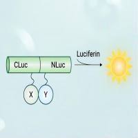
荧火素酶互补实验(Luciferase Complementation Assay, LCA)| 荧光素酶互补成像技术(Luciferase Complementation Imaging, LCI)
¥5999

UltraBio™ Mouse IgG Magnetic Beads (小鼠IgG磁珠),阿拉丁
¥932.90

