In Situ Proximity Ligation Assay for Microscopy and Flow Cytometry
互联网
- Abstract
- Table of Contents
- Materials
- Figures
- Literature Cited
Abstract
The ability to study proteins and protein interactions inside cells and tissues is important for elucidating how cells function in health and disease. The in situ proximity ligation assay (in situ PLA) presented here can be used to visualize proteins, protein?protein interactions, and post?translational modifications in cells and tissues. The method is based upon the use of antibodies that target the proteins involved in an interaction; hence, the method has the advantage that it can be used in clinical specimens, providing localized, quantifiable single molecule detection in single cells. This unit describes how in situ PLA can be used with fluorescence microscopy and flow cytometry to study proteins (obtaining high sensitivity and specificity of detection) and protein interactions. It also includes information on expected results and information on how to troubleshoot the assay. Curr. Protoc. Cytom. 56:9.36.1?9.36.15. © 2011 by John Wiley & Sons, Inc.
Keywords: in situ; proximity ligation; protein interactions; post?translational modifications
Table of Contents
- Introduction
- Basic Protocol 1: In Situ PLA for Microscopy
- Basic Protocol 2: In Situ PLA for Flow Cytometry
- Support Protocol 1: Conjugation of Oligonucleotides to Antibodies
- Reagents and Solutions
- Commentary
- Literature Cited
- Figures
- Tables
Materials
Basic Protocol 1: In Situ PLA for Microscopy
Materials
Basic Protocol 2: In Situ PLA for Flow Cytometry
Materials
Support Protocol 1: Conjugation of Oligonucleotides to Antibodies
Materials
|
Figures
-
Figure 9.36.1 Schematic overview of the steps involved with in situ PLA. The process consists of (A ) binding of the proximity probes to the target proteins, (B ) hybridization and joining of two linear connector oligonucleotides into a DNA circle, (C ) rolling circle amplification of the DNA circle, and (D ) hybridization of fluorescent detection oligonucleotides to the repeated sequences of the rolling circle product. View Image -
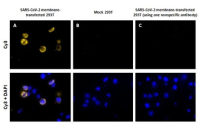
Figure 9.36.2 Typical in situ PLA results for sensitive detection of a protein. Detection of the HER2 receptor in (A ) MCF‐7 cells with a low expression level of the protein, or in (B ) SKOV3 cells with a high expression level. Upon successful detection of the protein, a fluorescent spot (red) is formed through the in situ PLA reaction. However, when the expression level of the detected protein is too high, the spots can start to coalesce (B). In situ PLA was performed with secondary proximity probes targeting a mouse monoclonal anti‐HER2 antibody and a rabbit polyclonal anti‐HER2 antibody. Cell nuclei were counterstained with Hoechst dye (blue). View Image -
Figure 9.36.3 Interactions between EGFR and HER2 in a formalin‐fixed paraffin‐embedded tissue section from a breast cancer patient is detected by in situ PLA. Antibodies targeting EGFR and HER2 were used in combination with secondary proximity probes. Red dots indicate detected protein‐protein interactions, blue indicates cell nuclei stained with Hoechst dye, and green is the autofluorescence in the FITC channel used to visualize the cytoplasm. View Image
Videos
Literature Cited
| Literature Cited | |
| Allalou, A. and Wählby, C. 2009. BlobFinder, a tool for fluorescence microscopy image cytometry. Comput. Methods Programs Biomed. 94:58‐65. | |
| Baan, B., Pardali, E., ten Dijke, P., and van Dam, H. 2010. In situ proximity ligation detection of c‐Jun/AP‐1 dimers reveals increased levels of c‐Jun/Fra1 complexes in aggressive breast cancer cell lines in vitro and in vivo. Mol. Cell Proteomics 9:1982‐1990. | |
| Carpenter, A.E., Jones, T.R., Lamprecht, M.R., Clarke, C., Kang, I.H., Friman, O., Guertin, D.A., Chang, J.H., Lindquist, R.A., Moffat, J., Golland, P., and Sabatini, D.M. 2006. CellProfiler: Image analysis software for identifying and quantifying cell phenotypes. Genome Biol. 7:R100. | |
| Frolova, E.I., Gorchakov, R., Pereboeva, L., Atasheva, S., and Frolov, I. 2010. Functional Sindbis virus replicative complexes are formed at the plasma membrane. J. Virol. 84:11679‐11695. | |
| Hu, C.D., Chinenov, Y., and Kerppola, T.K. 2002. Visualization of interactions among bZIP and Rel family proteins in living cells using bimolecular fluorescence complementation. Mol. Cell 9:789‐798. | |
| Jarvius, M., Paulsson, J., Weibrecht, I., Leuchowius, K.J., Andersson, A.C., Wählby, C., Gullberg, M., Botling, J., Sjöblom, T., Markova, B., Östman, A., Landegren, U., and Söderberg, O. 2007. In situ detection of phosphorylated platelet‐derived growth factor receptor beta using a generalized proximity ligation method. Mol. Cell Proteomics 6:1500‐1509. | |
| Johansson, H., Svensson, F., Runnberg, R., Simonsson, T., and Simonsson, S. 2010. Phosphorylated nucleolin interacts with translationally controlled tumor protein during mitosis and with Oct4 during interphase in ES cells. PLoS One 5:e13678. | |
| Kenworthy, A.K. 2001. Imaging protein‐protein interactions using fluorescence resonance energy transfer microscopy. Methods 24:289‐296. | |
| Lasserre, R., Charrin, S., Cuche, C., Danckaert, A., Thoulouze, M.I., de Chaumont, F., Duong, T., Perrault, N., Varin‐Blank, N., Olivo‐Marin, J.C., Etienne‐Manneville, S., Arpin, M., Di Bartolo, V., and Alcover, A. 2010. Ezrin tunes T‐cell activation by controlling Dlg1 and microtubule positioning at the immunological synapse. EMBO J. 29:2301‐2314. | |
| Leuchowius, K.J., Weibrecht, I., Landegren, U., Gedda, L., and Söderberg, O. 2009. Flow cytometric in situ proximity ligation analyses of protein interactions and post‐translational modification of the epidermal growth factor receptor family. Cytometry A 75:833‐839. | |
| Leuchowius, K.J., Jarvius, M., Wickström, M., Rickardson, L., Landegren, U., Larsson, R., Söderberg, O., Fryknas, M., and Jarvius, J. 2010. High content screening for inhibitors of protein interactions and post‐translational modifications in primary cells by proximity ligation. Mol. Cell Proteomics 9:178‐183. | |
| Sehat, B., Tofigh, A., Lin, Y., Trocme, E., Liljedahl, U., Lagergren, J., and Larsson, O. 2010. SUMOylation mediates the nuclear translocation and signaling of the IGF‐1 receptor. Sci. Signal 3:ra10. | |
| Söderberg, O., Gullberg, M., Jarvius, M., Ridderstråle, K., Leuchowius, K.J., Jarvius, J., Wester, K., Hydbring, P., Bahram, F., Larsson, L.G., and Landegren, U. 2006. Direct observation of individual endogenous protein complexes in situ by proximity ligation. Nat. Methods 3:995‐1000. |